. 2011 Jun 24;286(25):22243-9.
doi: 10.1074/jbc.M111.225755. Epub 2011 May 4.

First structural glimpse of CCN3 and CCN5 multifunctional signaling regulators elucidated by small angle x-ray scattering

Affiliations

First structural glimpse of CCN3 and CCN5 multifunctional signaling regulators elucidated by small angle x-ray scattering

Kenneth P Holbourn et al. J Biol Chem. .

Abstract

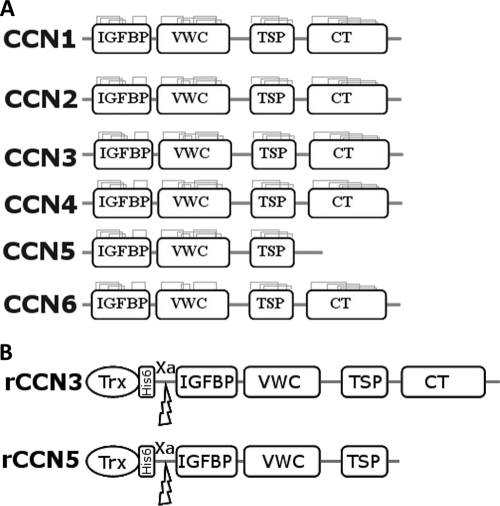
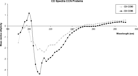
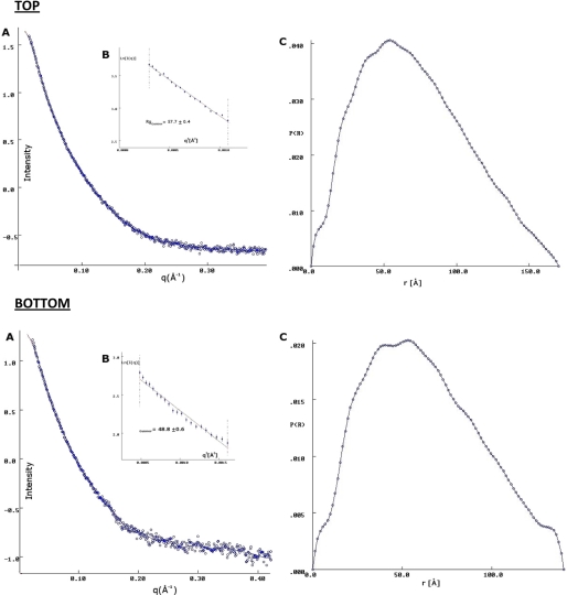
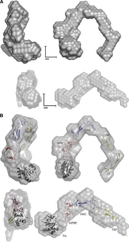
The CCN (cyr61, ctgf, nov) proteins (CCN1-6) are an important family of matricellular regulatory factors involved in internal and external cell signaling. They are central to essential biological processes such as adhesion, proliferation, angiogenesis, tumorigenesis, wound healing, and modulation of the extracellular matrix. They possess a highly conserved modular structure with four distinct modules that interact with a wide range of regulatory proteins and ligands. However, at the structural level, little is known although their biological function(s) seems to require cooperation between individual modules. Here we present for the first time structural determinants of two of the CCN family members, CCN3 and CCN5 (expressed in Escherichia coli), using small angle x-ray scattering. The results provide a description of the overall molecular shape and possible general three-dimensional modular arrangement for CCN proteins. These data unequivocally provide insight of the nature of CCN protein(s) in solution and thus important insight into their structure-function relationships.


Figures

FIGURE 1.
FIGURE 1.
A schematic diagram of the CCN family. A, the six members of the CCN family illustrating the four conserved domains. The conserved cysteine bonds are also shown in each domain with CCN6 lacking two of these in the VWC domain. CCN5 can also be seen to be lacking the CT domain (6). B, a schematic of the recombinant (r) constructs of CCN3 and CCN5 with an N-terminal Trx tag, an N-terminal hexa-histidine affinity tag, and a factor Xa cleavage site.
FIGURE 2.
FIGURE 2.
Circular dichroism data. CD spectra of CCN3 (gray triangles) and CCN5 (black squares) are shown. CD spectra were collected on a Jasco-J600 between 190 and 290 nm. Analysis in the program JFIT suggested that ∼50% of the protein was composed of β-strands, ∼40% consisted of random coil, and ∼10% consisted of α-helix. The large proportion of β-strand and coil is consistent with the homology modeling with the small proportion of α-helix likely from the Trx tag.
FIGURE 3.
FIGURE 3.
Scattering curves for CCN3 and CCN5. Top, A, the scattering curve of CCN3 (blue circles). The fit to the ab initio data is shown in red and was calculated using the program DAMMIN (41). B, the radius of gyration (Rg) was calculated using the Guinier approximation (61) and was found to be 57.7 Å. C, the interatomic distance function (P(r)) gave a maximum dimension of 170 Å. These values were calculated in GNOM (44). Bottom, A, the scattering curve of CCN5 (blue circles). The fit to the ab initio data is shown in red and was calculated using the program DAMMIN (41). B, the radius of gyration (Rg) was calculated using the Guinier approximation (61) and was found to be 48.8 Å. C, the interatomic distance function (P(r)) gave a maximum dimension of 160 Å. These values were calculated in GNOM (44).
FIGURE 4.
FIGURE 4.
Model fitting to small angle x-ray scattering data. A, the ab initio models generated by DAMMIN (45) for CCN3 and CCN5. In both cases, they are long elongated molecules that are extended and may act as a scaffold for binding and coordinating ligand partners. B, the homology models generated of the individual domains of the CCN proteins modeled into the surface envelope of the small angle x-ray scattering model. The domains are colored with the Trx tag in black, the IGFBP domain in red, the VWC domain in blue, the TSP domain in yellow, and the CT domain in green. CCN5 lacks the CT domain. The figures were generated using PyMOL (62).

Similar articles

Cited by

References

    1. Holbourn K. P., Acharya K. R., Perbal B. (2008) Trends Biochem. Sci. 33, 461–473 - PMC - PubMed
    1. Holbourn K. P., Perbal B., Acharya K. R. (2009) J. Cell Commun. Signal. 3, 25–41 - PMC - PubMed
    1. O'Brien T. P., Yang G. P., Sanders L., Lau L. F. (1990) Mol. Cell. Biol. 10, 3569–3577 - PMC - PubMed
    1. Bradham D. M., Igarashi A., Potter R. L., Grotendorst G. R. (1991) J. Cell Biol. 114, 1285–1294 - PMC - PubMed
    1. Joliot V., Martinerie C., Dambrine G., Plassiart G., Brisac M., Crochet J., Perbal B. (1992) Mol. Cell. Biol. 12, 10–21 - PMC - PubMed

Publication types

MeSH terms

Substances

LinkOut - more resources