. 2011 Jul 29;286(30):26718-31.
doi: 10.1074/jbc.M111.244590. Epub 2011 May 4.

N-formyl peptide receptor 3 (FPR3) departs from the homologous FPR2/ALX receptor with regard to the major processes governing chemoattractant receptor regulation, expression at the cell surface, and phosphorylation

Affiliations

N-formyl peptide receptor 3 (FPR3) departs from the homologous FPR2/ALX receptor with regard to the major processes governing chemoattractant receptor regulation, expression at the cell surface, and phosphorylation

Marie-Josèphe Rabiet et al. J Biol Chem. .

Abstract

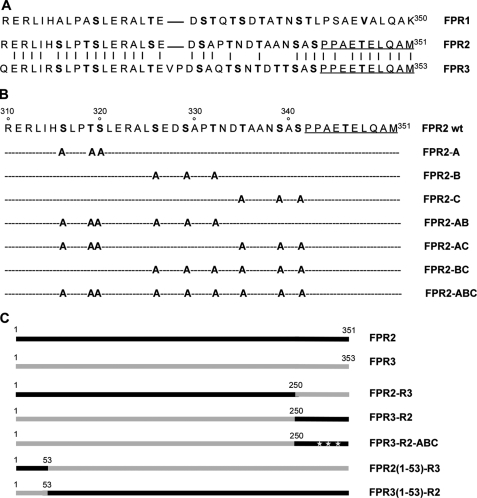
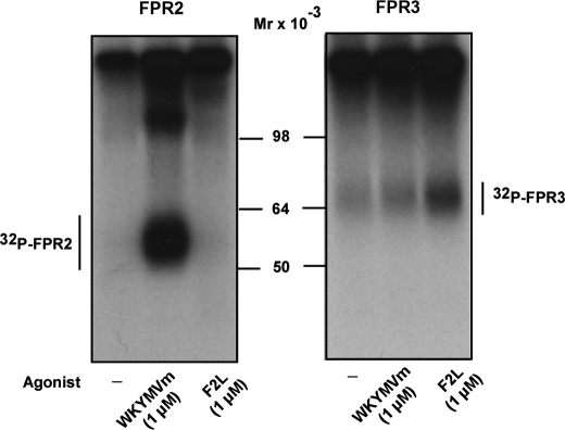
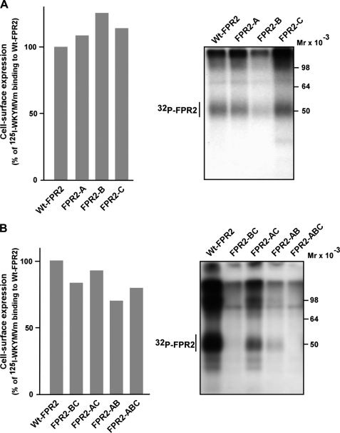
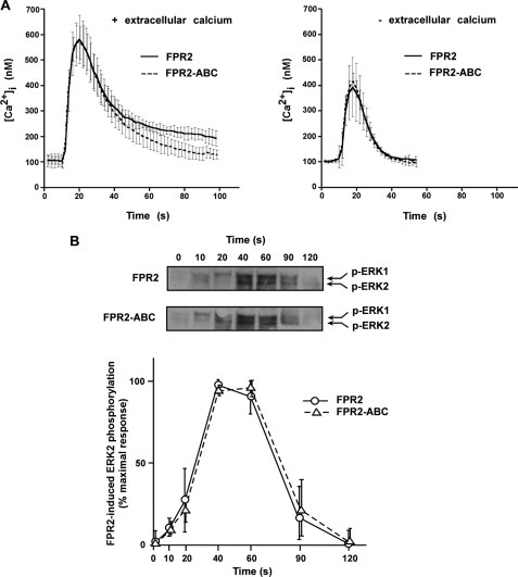
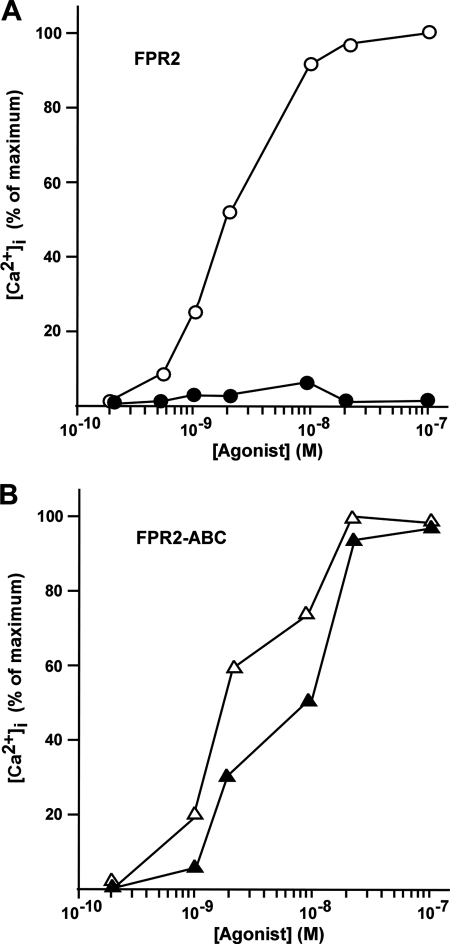
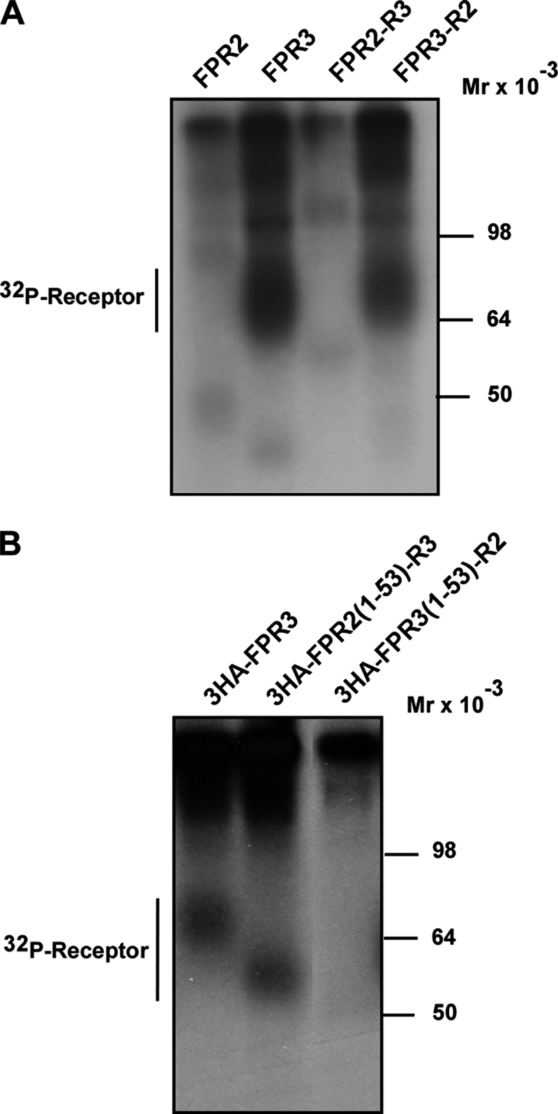
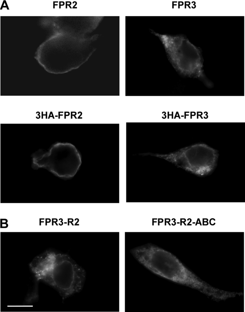
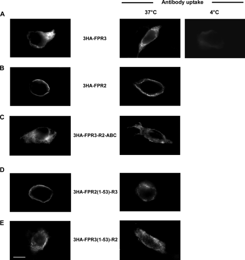
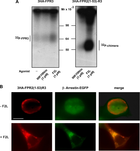
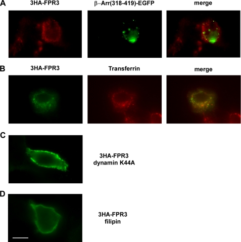
Among human N-formyl peptide chemoattractant receptors, FPR2/ALX and FPR3 share the highest degree of amino acid identity (83%), and trigger similar cell responses upon ligand binding. Although FPR2/ALX is a promiscuous receptor, FPR3 has only one specific high affinity ligand, F2L, and a more restricted tissue/cell distribution. In this study, we showed that FPR2/ALX behaved as the prototypical receptor FPR1. The agonist-dependent phosphorylation used a hierarchical mechanism with a prominent role of Ser(329), Thr(332), and Thr(335). Phosphorylation of FPR2/ALX was essential for its desensitization but the lack of phosphorylation did not result in enhanced or sustained responses. In contrast, resting FPR3 displayed a marked level of phosphorylation, which was only slightly increased upon agonist stimulation. Another noticeable difference between the two receptors was their subcellular distribution in unstimulated cells. Although FPR2/ALX was evenly distributed at the plasma membrane FPR3 was localized in small intracellular vesicles. By swapping domains between FPR2/ALX and FPR3, we uncovered the determinants involved in the basal phosphorylation of FPR3. Experiments aimed at monitoring receptor-bound antibody uptake showed that the intracellular distribution of FPR3 resulted from a constitutive internalization that was independent of C terminus phosphorylation. Unexpectedly, exchanging residues 1 to 53, which encompass the N-terminal extracellular region and the first transmembrane domain, between FPR2/ALX and FPR3 switched localization of the receptors from the plasma membrane to intracellular vesicles and vice versa. A clathrin-independent, possibly caveolae-dependent, mechanism was involved in FPR3 constitutive internalization. The peculiar behavior of FPR3 most probably serves distinct physiological functions that remain largely unknown.


Figures

FIGURE 1.
FIGURE 1.
Schematic representation of mutant and chimeric receptors. A, alignment of the primary sequence of the intracellular carboxyl terminus of human FPR1, FPR2/ALX, and FPR3. Residues that are identical in FPR2/ALX and FPR3 are linked by a vertical hyphen. B, amino acid sequence of the carboxyl terminus of wild type FPR2/ALX and location of point mutations in FPR2/ALX. Positions of putative phosphorylation sites in the primary sequence of the wild type FPR2/ALX C terminus are indicated in bold. Altered residues are shown for the different mutants. Residues corresponding to the anti-FPR2 epitope are underlined. C, schematic representation of wild type and chimeric receptors obtained by swapping C-terminal or N-terminal domains. The topology of FPR2/ALX (black lines) and FPR3 (gray lines) in chimeric receptors is illustrated.
FIGURE 2.
FIGURE 2.
Agonist-induced phosphorylation of FPR2/ALX and FPR3. HEK293T cells expressing wild type FPR2/ALX (right panel) or FPR3 (left panel) were metabolically labeled with [32P]orthophosphoric acid and incubated for 15 min at 37 °C with no agonist (first lane), 1 μm WKYMVm (second lane), or 1 μm F2L (third lane). After cell lysis, receptors were immunoprecipitated with an antibody directed against the last 10 amino acids of FPR2/ALX that cross-reacts with FPR3. Immunoprecipitates were treated with 2-fold Laemmli sample buffer and subjected to SDS-PAGE and autoradiography. Data are representative of three independent experiments.
FIGURE 3.
FIGURE 3.
Cell surface expression and phosphorylation of the wild type and mutant forms of FPR2/ALX. A, cell surface expression. HEK293T cells expressing wild type or mutant FPR2/ALX were incubated with increasing concentrations of 125I-labeled WKYMVm peptide for 1 h at 4 °C. Radioactivity specifically bound to cells was recorded with a γ-counter. The results are expressed as the percentage of 125I-labeled peptide bound on wild type FPR2/ALX expressing cells. B, receptor phosphorylation. HEK293T cells that expressed wild type or mutant forms of FPR2/ALX were metabolically labeled with [32P]orthophosphoric acid. Cells were stimulated with 100 nm WKYMVm for 15 min at 37 °C. Receptors were immunoprecipitated from cell lysates with an antibody directed against the last 10 amino acids of FPR2/ALX. Receptor immunoprecipitates were resolved by SDS-PAGE and analyzed by autoradiography. Data are representative of three independent experiments.
FIGURE 4.
FIGURE 4.
Agonist-induced intracellular signaling by the wild type FPR2/ALX and mutant FPR2-ABC. A, agonist-induced calcium mobilization. HL60 cells expressing the wild type (solid line) and mutant form of FPR2/ALX (dashed line) were analyzed for agonist-induced desensitization of calcium mobilization. Following loading with Fura2, cells were stimulated with 100 nm WKYMVm and assayed for calcium mobilization in the presence (left panel) or absence (right panel) of extracellular calcium. Data are expressed as mean responses ± S.E. (n > 3). B, agonist-induced ERK1/2 phosphorylation. HL60 cells expressing the wild type FPR2/ALX and the mutant FPR2-ABC were stimulated with 100 nm WKYMVm for various periods of time. Phosphorylation of ERK1/2 was visualized by immunoblotting with anti-phospho-ERK1/2 antibody (upper panel). Blots are representative of three experiments. Western blot analyses with an anti-ERK2 antibody were performed to check that an equal amount of ERK2 was present in each sample (not shown). Phosphorylation of ERK2 was subsequently quantified by densitometry (lower panel). The results are expressed in percent of maximum phosphorylation in wild type expressing cells. Values in graphs represent the mean ± S.E. from three independent experiments.
FIGURE 5.
FIGURE 5.
Desensitization of calcium mobilization by wild type FPR2/ALX and phosphorylation-deficient mutant FPR2-ABC. HL60 cells expressing FPR2/ALX (A) and FPR2-ABC (B) were analyzed for agonist-induced desensitization of calcium mobilization. Fura-2-loaded cells were incubated with either 100 nm WKYMVm (black symbols) or vehicle (clear symbols) for 10 min at 37 °C. Cells were then washed three times at room temperature and subsequently assayed for calcium mobilization in response to various doses of WKYMVm. Data expressed in percent of the maximum response are representative of three independent experiments.
FIGURE 6.
FIGURE 6.
Constitutive phosphorylation of FPR3 and chimeric receptors obtained by swapping C-terminal or N-terminal domains. HEK293 cells expressing either FPR3 or the indicated chimeric receptors were metabolically labeled with [32P]orthophosphoric acid. Cells were not challenged with agonist. Receptors were immunoprecipitated from cell lysates with the anti-FPR2/ALX antibody (A) or with an anti-HA antibody when cells expressed 3HA-tagged receptors (B). Receptor immunoprecipitates were resolved by SDS-PAGE and analyzed by autoradiography. Data are representative of three independent experiments.
FIGURE 7.
FIGURE 7.
Cellular distribution of FPR2, FPR3, and chimeric receptors FPR3-R2 and FPR3-R2-ABC. HEK293 cells were transiently transfected with FPR2/ALX or FPR3 (tagged or not with 3HA) (A), or the chimeric receptors obtained by swapping C-terminal domains (B). Cells were fixed and permeabilized. The cellular distribution of receptors was visualized by labeling with either a polyclonal anti-FPR2/ALX antibody, which cross-react with FPR3, or a monoclonal anti-HA antibody followed by immunofluorescence staining with Alexa Fluor 488-conjugated secondary antibodies. Scale bar, 10 μm.
FIGURE 8.
FIGURE 8.
Constitutive internalization of FPR3. Antibody uptake (right panels) was performed on living cells. Cells expressing the indicated HA-tagged receptors were incubated with a monoclonal anti-HA antibody for 30 min at 37 °C (or at 4 °C to inhibit internalization) to label the receptors present on the cell surface. Subsequently, cells were fixed, permeabilized, incubated with anti-mouse Alexa Fluor 488-conjugated antibody, and processed for fluorescence microscopy. In parallel (left panels), the distribution of the receptors was visualized in cells fixed and permeabilized prior to incubation with monoclonal anti-HA antibody and immunofluorescence staining. Scale bar, 10 μm.
FIGURE 9.
FIGURE 9.
Agonist-induced phosphorylation and internalization of chimeric receptors. A, HEK293 cells expressing 3HA-tagged FPR3 or the chimeric receptor in which the N-terminal domain of FPR2/ALX and FPR3 has been exchanged (3HA-FPR2(1–53)-R3), were metabolically labeled with [32P]orthophosphoric acid. Cells were not stimulated or stimulated for 15 min at 37 °C with 1 μm WKYMVm or 1 μm F2L as indicated. After cell lysis, receptors were immunoprecipitated with an anti-HA antibody. Receptor immunoprecipitates were resolved by SDS-PAGE and analyzed by autoradiography. Data are representative of two independent experiments. B, HEK293 cells were cotransfected with the chimera 3HA-FPR2(1–53)-R3 and β-arrestin-1 in fusion with EGFP. Cells were not stimulated or stimulated with 1 μm F2L for 30 min, at 37 °C. Cells were fixed and permeabilized. The 3HA-FPR2(1–53)-R3 receptor was visualized by incubation with monoclonal anti-HA antibody and staining with a red fluorescent Alexa Fluor 568-conjugated anti-mouse antibody. Scale bar, 10 μm.
FIGURE 10.
FIGURE 10.
Endocytosis pathway involved in FPR3 constitutive internalization. A, the 3HA-FPR3 receptor was coexpressed in HEK293 cells with a fragment of β-arrestin 1 (amino acids 318–419) in fusion with EGFP (β-Arr(318–419)-EGFP). The 3HA-FPR3 was labeled with the monoclonal anti-HA and a red fluorescent Alexa 568-conjugated anti-mouse antibody. B, antibody uptake experiments were performed on living cells expressing 3HA-FPR3. The cells were incubated with anti-HA antibody for 30 min at 37 °C. Internalization of the transferrin receptor was visualized by adding transferrin-Alexa Fluor 568 conjugate during the last 15 min of incubation. Cells were fixed, permeabilized, and incubated with an Alexa Fluor 488-conjugated anti-mouse antibody to label the anti-HA antibody bound to the internalized 3HA-FPR3. C, antibody uptake was performed in HEK293 cells cotransfected with 3HA-FPR3 and the dominant-negative mutant of dynamin (dynamin K44A). D, antibody uptake was performed in HEK293 cells expressing the 3HA-FPR3 receptor in the presence of filipin III. Scale bar, 10 μm.

Similar articles

Cited by

References

    1. Ye R. D., Boulay F., Wang J. M., Dahlgren C., Gerard C., Parmentier M., Serhan C. N., Murphy P. M. (2009) Pharmacol. Rev. 61, 119–161 - PMC - PubMed
    1. Rabiet M. J., Huet E., Boulay F. (2005) Eur. J. Immunol. 35, 2486–2495 - PubMed
    1. Christophe T., Karlsson A., Dugave C., Rabiet M. J., Boulay F., Dahlgren C. (2001) J. Biol. Chem. 276, 21585–21593 - PubMed
    1. Migeotte I., Riboldi E., Franssen J. D., Grégoire F., Loison C., Wittamer V., Detheux M., Robberecht P., Costagliola S., Vassart G., Sozzani S., Parmentier M., Communi D. (2005) J. Exp. Med. 201, 83–93 - PMC - PubMed
    1. McDonald P. H., Lefkowitz R. J. (2001) Cell. Signal. 13, 683–689 - PubMed

LinkOut - more resources