. 2011 Jun 17;286(24):21937-43.
doi: 10.1074/jbc.M111.230706. Epub 2011 May 4.

Structure of the autocatalytic cysteine protease domain of potyvirus helper-component proteinase

Affiliations

Structure of the autocatalytic cysteine protease domain of potyvirus helper-component proteinase

Bihong Guo et al. J Biol Chem. .

Abstract

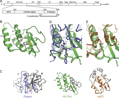
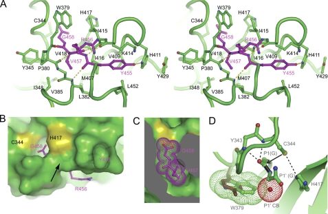
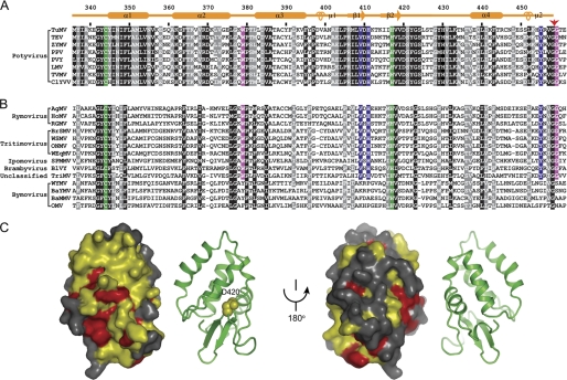
The helper-component proteinase (HC-Pro) of potyvirus is involved in polyprotein processing, aphid transmission, and suppression of antiviral RNA silencing. There is no high resolution structure reported for any part of HC-Pro, hindering mechanistic understanding of its multiple functions. We have determined the crystal structure of the cysteine protease domain of HC-Pro from turnip mosaic virus at 2.0 Å resolution. As a protease, HC-Pro only cleaves a Gly-Gly dipeptide at its own C terminus. The structure represents a postcleavage state in which the cleaved C terminus remains tightly bound at the active site cleft to prevent trans activity. The structure adopts a compact α/β-fold, which differs from papain-like cysteine proteases and shows weak similarity to nsP2 protease from Venezuelan equine encephalitis alphavirus. Nevertheless, the catalytic cysteine and histidine residues constitute an active site that is highly similar to these in papain-like and nsP2 proteases. HC-Pro recognizes a consensus sequence YXVGG around the cleavage site between the two glycine residues. The structure delineates the sequence specificity at sites P1-P4. Structural modeling and covariation analysis across the Potyviridae family suggest a tryptophan residue accounting for the glycine specificity at site P1'. Moreover, a surface of the protease domain is conserved in potyvirus but not in other genera of the Potyviridae family, likely due to extra functional constrain. The structure provides insight into the catalysis mechanism, cis-acting mode, cleavage site specificity, and other functions of the HC-Pro protease domain.


Figures

FIGURE 1.
FIGURE 1.
Structure of HC-Pro CPD from TuMV. A, diagram of the TuMV RNA genome, the domain organization of HC-Pro, and the construct used for crystallization. Mature products for the polyprotein are indicated. B, ribbon representation of the CPD structure. The secondary structural elements and the N and C terminus are indicated. The catalytic dyad residues Cys344 and His417 (yellow) and the C-terminal residue Gly458 (magenta) are shown as sticks. C, structural comparison of HC-Pro CPD, papain (Protein Data Bank ID code 6PAD), and nsP2 CPD of Venezuelan equine encephalitis alphavirus (Protein Data Bank ID code 2HWK). The three structures are aligned around the catalytic dyad residues. The equivalent structure elements are colored green in HC-Pro, blue in papain, and orange in nsP2, whereas the rest of the structures are gray. The catalytic residues and substrate/inhibitor are shown in sticks and are colored in yellow and magenta, respectively. D and E, superposition of the active site of HC-Pro CPD with that of papain covalently linked to an inhibitor (D) and nsP2 (E).
FIGURE 2.
FIGURE 2.
Recognition of the cleavage site. A, Cross-eye stereo view of the C-terminal tetrapeptide (P1–P4) bound at the active site cleft. Interacting residues are shown in stick-and-ball format. Oxygen atoms are red, nitrogen atoms are blue, and carbon atoms are magenta in the tetrapeptide and green for other residues. Dashed lines denote hydrogen bonds. B, surface view of the active site cleft with the bound C-terminal tetrapeptide. C, cut-away view of the S1 and S2 pockets. The P1 residue Gly458 and P2 residue Val457 are shown as dots. D, structural model of the precleavage state. The side chains of the catalytic dyad residues are reoriented to mimic an active configuration, in which Cys344 is deprotonated by His417 and acts as a nucleophile to attack the carbonyl carbon of the scissile bond (black). The amides of Cys344 and Tyr343 may constitute an oxyanion hole to stabilize the transition intermediate. If P1′ has a CB atom (red dots), the CB atom would clash with Trp379 (green dots). Replacement of Trp379 by tyrosine (brown) observed in Bymovirus may remove the specificity for P1′ glycine.
FIGURE 3.
FIGURE 3.
Sequence conservation in the HC-Pro CPD. A, alignment of sequences from the Potyvirus genus. The black and gray shading represent 100 and 90% sequence similarity in 59 aligned sequences, respectively, eight of which are displayed. Similarity groups are defined as follows: S and T, D and E, K and R, and L, I, V, M, F, Y, and W. The secondary structure elements and residue numbers are indicated for TuMV HC-Pro. The red arrow points to the self-cleavage site. The catalytic dyad residues are shaded in green. The P1′ residue Gly458, the putative S1′ residue Trp379 and their equivalents that are invariant in other sequences are shaded in magenta. The P4 residue Tyr455, the S4 residues Val409 and His411 and their equivalents invariant in other sequences are shaded in yellow. ZYMV, zucchini yellow mosaic virus; PPV, plum pox virus; PVY, potato virus Y; LMV, lettuce mosaic virus; TVMV, tobacco vein mottling virus; ClYVV, clover yellow vein virus. B, conservation in the Potyviridae family. Five potyviral HC-Pro proteins (TuMV, TEV, ZYMV, PPV, and PVY, not shown) and all available homologs from other genera of the Potyviridae family are aligned. Residues with 100 and 90% conservation in these sequences are shaded in black and gray, respectively. The genera are indicated. AgMV, agropyron mosaic virus; HoMV, hordeum mosaic virus; RGMV, ryegrass mosaic virus, BrSMV, brome streak mosaic virus; WSMV, wheat streak mosaic virus; ONMV, oat necrotic mottle virus; WEqMV, wheat eqlid mosaic virus; SPMMV, sweet potato mild mottle virus; BlVY, blackberry virus Y; TriMV, Triticum mosaic virus; WYMV, wheat yellow mosaic virus; BaYMV, barley yellow mosaic virus; BaMMV, barley mild mosaic virus; OMV, oat mosaic virus. C, conserved surface at two opposite orientations. Residues at least 90% conserved in the genus Potyvirus as defined in A are colored yellow, and those at least 90% conserved across the Potyviridae family as defined in B are colored red. The corresponding ribbon representations are shown in parallel. Residue Asp420 is shown as spheres.

Similar articles

Cited by

References

    1. Dougherty W. G., Semler B. L. (1993) Microbiol. Rev. 57, 781–822 - PMC - PubMed
    1. Urcuqui-Inchima S., Haenni A. L., Bernardi F. (2001) Virus Res. 74, 157–175 - PubMed
    1. Verchot J., Koonin E. V., Carrington J. C. (1991) Virology 185, 527–535 - PubMed
    1. Mavankal G., Rhoads R. E. (1991) Virology 185, 721–731 - PubMed
    1. Carrington J. C., Cary S. M., Parks T. D., Dougherty W. G. (1989) EMBO J. 8, 365–370 - PMC - PubMed

Publication types

Associated data