Identification of dihydroceramide desaturase as a direct in vitro target for fenretinide
- PMID: 21543327
- PMCID: PMC3137051
- DOI: 10.1074/jbc.M111.250779
Identification of dihydroceramide desaturase as a direct in vitro target for fenretinide
Abstract
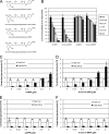
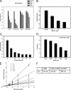
The dihydroceramide desaturase (DES) enzyme is responsible for inserting the 4,5-trans-double bond to the sphingolipid backbone of dihydroceramide. We previously demonstrated that fenretinide (4-HPR) inhibited DES activity in SMS-KCNR neuroblastoma cells. In this study, we investigated whether 4-HPR acted directly on the enzyme in vitro. N-C8:0-d-erythro-dihydroceramide (C(8)-dhCer) was used as a substrate to study the conversion of dihydroceramide into ceramide in vitro using rat liver microsomes, and the formation of tritiated water after the addition of the tritiated substrate was detected and used to measure DES activity. NADH served as a cofactor. The apparent K(m) for C(8)-dhCer and NADH were 1.92 ± 0.36 μm and 43.4 ± 6.47 μm, respectively; and the V(max) was 3.16 ± 0.24 and 4.11 ± 0.18 nmol/min/g protein. Next, the effects of 4-HPR and its metabolites on DES activity were investigated. 4-HPR was found to inhibit DES in a dose-dependent manner. At 20 min, the inhibition was competitive; however, longer incubation times demonstrated the inhibition to be irreversible. Among the major metabolites of 4-HPR, 4-oxo-N-(4-hydroxyphenyl)retinamide (4-oxo-4-HPR) showed the highest inhibitory effect with substrate concentration of 0.5 μm, with an IC(50) of 1.68 μm as compared with an IC(50) of 2.32 μm for 4-HPR. N-(4-Methoxyphenyl)retinamide (4-MPR) and 4-Oxo-N-(4-methoxyphenyl)retinamide (4-oxo-4-MPR) had minimal effects on DES activity. A known competitive inhibitor of DES, C(8)-cyclopropenylceramide was used as a positive control. These studies define for the first time a direct in vitro target for 4-HPR and suggest that inhibitors of DES may be used as therapeutic interventions to regulate ceramide desaturation and consequent function.
Figures


Similar articles
-
Inhibitory effects of fenretinide metabolites N-[4-methoxyphenyl]retinamide (MPR) and 4-oxo-N-(4-hydroxyphenyl)retinamide (3-keto-HPR) on fenretinide molecular targets β-carotene oxygenase 1, stearoyl-CoA desaturase 1 and dihydroceramide Δ4-desaturase 1.PLoS One. 2017 Apr 27;12(4):e0176487. doi: 10.1371/journal.pone.0176487. eCollection 2017. PLoS One. 2017. PMID: 28448568 Free PMC article.
-
Involvement of dihydroceramide desaturase in cell cycle progression in human neuroblastoma cells.J Biol Chem. 2007 Jun 8;282(23):16718-28. doi: 10.1074/jbc.M700647200. Epub 2007 Feb 5. J Biol Chem. 2007. PMID: 17283068 Free PMC article.
-
Fenretinide metabolism in humans and mice: utilizing pharmacological modulation of its metabolic pathway to increase systemic exposure.Br J Pharmacol. 2011 Jul;163(6):1263-75. doi: 10.1111/j.1476-5381.2011.01310.x. Br J Pharmacol. 2011. PMID: 21391977 Free PMC article.
-
Clinical development of fenretinide as an antineoplastic drug: Pharmacology perspectives.Exp Biol Med (Maywood). 2017 Jun;242(11):1178-1184. doi: 10.1177/1535370217706952. Epub 2017 Apr 21. Exp Biol Med (Maywood). 2017. PMID: 28429653 Free PMC article. Review.
-
The pathophysiological role of dihydroceramide desaturase in the nervous system.Prog Lipid Res. 2023 Jul;91:101236. doi: 10.1016/j.plipres.2023.101236. Epub 2023 May 13. Prog Lipid Res. 2023. PMID: 37187315 Review.
Cited by
-
Ceramide-orchestrated signalling in cancer cells.Nat Rev Cancer. 2013 Jan;13(1):51-65. doi: 10.1038/nrc3398. Epub 2012 Dec 13. Nat Rev Cancer. 2013. PMID: 23235911 Review.
-
Ceramide targets autophagosomes to mitochondria and induces lethal mitophagy.Nat Chem Biol. 2012 Oct;8(10):831-8. doi: 10.1038/nchembio.1059. Nat Chem Biol. 2012. PMID: 22922758 Free PMC article.
-
A Novel Nanomicellar Combination of Fenretinide and Lenalidomide Shows Marked Antitumor Activity in a Neuroblastoma Xenograft Model.Drug Des Devel Ther. 2019 Dec 19;13:4305-4319. doi: 10.2147/DDDT.S221909. eCollection 2019. Drug Des Devel Ther. 2019. PMID: 31908416 Free PMC article.
-
Sphingolipid Modulation Activates Proteostasis Programs to Govern Human Hematopoietic Stem Cell Self-Renewal.Cell Stem Cell. 2019 Nov 7;25(5):639-653.e7. doi: 10.1016/j.stem.2019.09.008. Epub 2019 Oct 17. Cell Stem Cell. 2019. PMID: 31631013 Free PMC article.
-
The Critical Impact of Sphingolipid Metabolism in Breast Cancer Progression and Drug Response.Int J Mol Sci. 2023 Jan 20;24(3):2107. doi: 10.3390/ijms24032107. Int J Mol Sci. 2023. PMID: 36768427 Free PMC article. Review.
References
-
- Hannun Y. A., Obeid L. M. (2008) Nat. Rev. Mol. Cell. Biol. 9, 139–150 - PubMed
-
- Ogretmen B., Hannun Y. A. (2004) Nat. Rev. Cancer 4, 604–616 - PubMed
-
- Wang H., Maurer B. J., Reynolds C. P., Cabot M. C. (2001) Cancer research 61, 5102–5105 - PubMed
-
- Michel C., van Echten-Deckert G., Rother J., Sandhoff K., Wang E., Merrill A. H., Jr. (1997) J. Biol. Chem. 272, 22432–22437 - PubMed
-
- Ternes P., Franke S., Zähringer U., Sperling P., Heinz E. (2002) J. Biol. Chem. 277, 25512–25518 - PubMed
Publication types
MeSH terms
Substances
Grants and funding
- P01-CA97132/CA/NCI NIH HHS/United States
- R01 CA087584/CA/NCI NIH HHS/United States
- R01-CA49837/CA/NCI NIH HHS/United States
- P01 CA097132/CA/NCI NIH HHS/United States
- K01-CA100767/CA/NCI NIH HHS/United States
- K01 CA100767/CA/NCI NIH HHS/United States
- P20 RR017677/RR/NCRR NIH HHS/United States
- R01-AG016583/AG/NIA NIH HHS/United States
- R01 CA049837/CA/NCI NIH HHS/United States
- C06 RR018823/RR/NCRR NIH HHS/United States
- C06-RR018823/RR/NCRR NIH HHS/United States
- R01 AG016583/AG/NIA NIH HHS/United States
- P20-RR17677/RR/NCRR NIH HHS/United States
LinkOut - more resources
Full Text Sources
Other Literature Sources

