. 2011 Jun 24;286(25):22469-77.
doi: 10.1074/jbc.M111.238469. Epub 2011 May 4.

The small GTPase Cdc42 is necessary for primary ciliogenesis in renal tubular epithelial cells

Affiliations

The small GTPase Cdc42 is necessary for primary ciliogenesis in renal tubular epithelial cells

Xiaofeng Zuo et al. J Biol Chem. .

Abstract

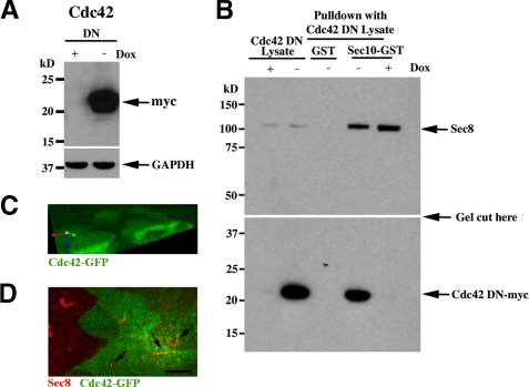
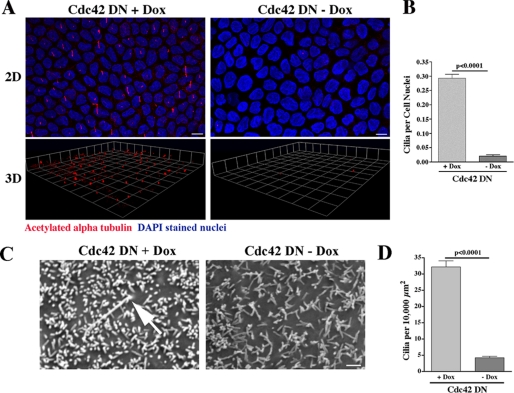
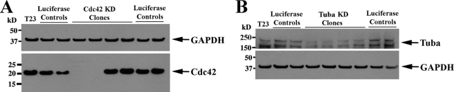
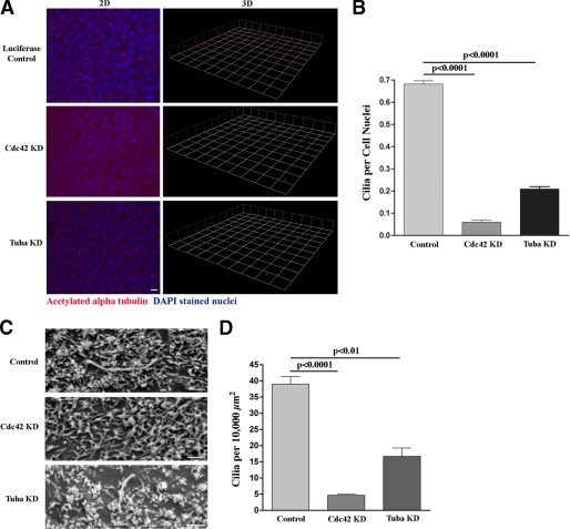
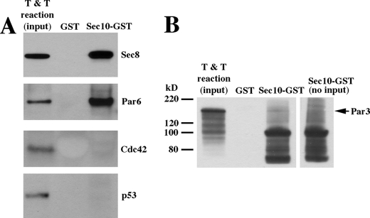
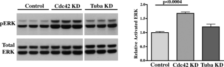
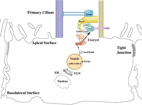
Primary cilia are found on many epithelial cell types, including renal tubular epithelial cells, where they participate in flow sensing. Disruption of cilia function has been linked to the pathogenesis of polycystic kidney disease. We demonstrated previously that the exocyst, a highly conserved eight-protein membrane trafficking complex, localizes to primary cilia of renal tubular epithelial cells, is required for ciliogenesis, biochemically and genetically interacts with polycystin-2 (the protein product of the polycystic kidney disease 2 gene), and, when disrupted, results in MAPK pathway activation both in vitro and in vivo. The small GTPase Cdc42 is a candidate for regulation of the exocyst at the primary cilium. Here, we demonstrate that Cdc42 biochemically interacts with Sec10, a crucial component of the exocyst complex, and that Cdc42 colocalizes with Sec10 at the primary cilium. Expression of dominant negative Cdc42 and shRNA-mediated knockdown of both Cdc42 and Tuba, a Cdc42 guanine nucleotide exchange factor, inhibit ciliogenesis in Madin-Darby canine kidney cells. Furthermore, exocyst Sec8 and polycystin-2 no longer localize to primary cilia or the ciliary region following Cdc42 and Tuba knockdown. We also show that Sec10 directly interacts with Par6, a member of the Par complex that itself directly interacts with Cdc42. Finally, we show that Cdc42 knockdown results in activation of the MAPK pathway, something observed in cells with dysfunctional primary cilia. These data support a model in which Cdc42 localizes the exocyst to the primary cilium, whereupon the exocyst then targets and docks vesicles carrying proteins necessary for ciliogenesis.


Figures

FIGURE 1.
FIGURE 1.
Exocyst Sec10 Interacts and Colocalizes with Cdc42. A, Western blot analysis of MDCK cells expressing dominant negative (DN) Cdc42, with a myc epitope tag, were grown in the presence or absence of doxycycline (Dox), a tetracycline derivative. In the absence of doxycycline, dominant negative Cdc42 was expressed. B, Sec10-GST fusion protein, generated and purified from E. coli., was incubated with MDCK cell lysate from Cdc42-myc DN mutant cells. After extensive washing, bound proteins were analyzed by Western blot analysis. DN Cdc42-myc was found in the pull-down fraction and, therefore, is a Sec10 binding partner. As a positive control for the Cdc42 DN lysates, Sec8 is shown to bind to Sec10-GST (top panel). As a negative control, GST alone was used, and no Cdc42 mutant protein was pulled down, even in the absence of doxycycline. The top and bottom panels are from the same gel, and the exposure for the top and bottom halves of the gel was the same. C, MDCK cells stably expressing Cdc42-GFP cells were grown on Transwell filters, and apical staining was seen as described previously (25). D, the Cdc42-GFP MDCK cells were fixed and stained with antibody against endogenous exocyst Sec8. Colocalization of Sec8 and Cdc42 is seen (arrows). Scale bar = 5 μm..
FIGURE 2.
FIGURE 2.
Dominant Negative Expression of Cdc42 Inhibits Ciliogenesis. A, MDCK cells expressing dominant negative (DN) Cdc42 in the presence and absence of doxycycline were grown on Transwell filters for 14 days. Using an antibody against acetylated α-tubulin (red), ciliogenesis was examined by confocal microscopy combined with three-dimensional (3D) reconstruction of the stacked series. Ciliogenesis was virtually completely inhibited when Cdc42 DN protein was expressed (in the absence of doxycycline (Dox)). DAPI (blue) is a nuclear stain. DAPI staining is at a different level in the cell than staining for acetylated α-tubulin but is included in the merged figure to delineate individual cells and allow for statistical analysis. Scale bar = 5 μm. B, quantification of ciliogenesis was performed using a ratio of cilia to cell nuclei. Significantly fewer cilia were seen in the MDCK cells expressing Cdc42 DN protein (-Dox). C, Cdc42 DN cells were grown on Transwell filters in the presence (+) and absence (-) of doxycycline for 14 days. The cells were fixed in glutaraldehyde, and SE microscopy was performed (Phillips XL20). Confirming the results in A, primary cilia were rarely seen when Cdc42 DN protein was expressed (-Dox). Scale bar = 1.0 μm. D, quantification of ciliogenesis was performed by counting the number of cilia per surface area, as individual cells could not be identified. Significantly fewer cilia were seen in the MDCK cells expressing Cdc42 DN protein (-Dox)..
FIGURE 3.
FIGURE 3.
Cdc42 and Tuba were significantly knocked down using shRNA. Lentivirus-encoding shRNA targeting Cdc42 (A) and Tuba (B) were used to infect MDCK cells (24). The lentiviral construct contained a GFP, which allowed us to use FACS to generate single cell clones. Stable knockdown cell lines were then grown, and Western blotting was performed to determine efficacy of knockdown. By Western blotting, there was complete knockdown of Cdc42 (A) and 70–80% knockdown of Tuba (B). Western blotting of GAPDH is shown to demonstrate equal protein loading in the gel lanes.
FIGURE 4.
FIGURE 4.
Knockdown of Cdc42 and Tuba inhibit ciliogenesis. A, MDCK cells containing stable shRNA-mediated knockdown of Cdc42 and Tuba were grown on Transwell filters for 14 days. Using an antibody against acetylated α-tubulin (red), ciliogenesis was examined by confocal microscopy combined with three-dimensional (3D) reconstruction of the stacked series. Ciliogenesis was virtually completely inhibited when Cdc42 was knocked down and significantly inhibited following Tuba knockdown. Again, please note that DAPI staining (blue) is at a different level in the cell than staining for acetylated α-tubulin but is included in the merged figure to delineate individual cells and allow for statistical analysis. Scale bar = 5 μm. B, quantification of ciliogenesis was performed using a ratio of cilia to cell nuclei. Significantly fewer cilia were seen in the MDCK cells following Cdc42 and Tuba knockdown. C, the same Cdc42 and Tuba knockdown MDCK cells were grown on Transwell filters for 14 days, fixed in glutaraldehyde, and then SE microscopy was performed (Phillips XL20). Confirming the results in (A), primary cilia were almost never seen following Cdc42 knockdown and were rarely seen following Tuba knockdown. Scale bar = 1.0 μm. D, quantification of ciliogenesis was again performed by counting the number of cilia per surface area, as individual cells could not be identified. Significantly fewer cilia were seen in the MDCK cells following Cdc42 and Tuba knockdown..
FIGURE 5.
FIGURE 5.
The Exocyst Directly Interacts with Par6. A, GST pull-downs using in vitro transcription and translation reactions (T & T) as input revealed that Par6 directly bound Sec10, whereas Cdc42 did not. We had previously shown by coimmunoprecipitation that the exocyst interacted with the Par complex (5), but we had not yet identified which member of the Par complex was directly binding to the exocyst. Sec10-GST pull-down of Sec8 is shown as a positive control, and p53, which is not known to bind the exocyst, is shown as a negative control. B, using in vitro transcription and translation reaction as input for a GST pull-down, the Par3-Sec10 interaction we previously showed by coimmunoprecipitation (5) is not detectable, indicating that the Par3-Sec10 interaction is indirect. The GST-Sec10 protein, at ∼100 kDa, gives some background on Western blot analyses, so we have included a control lane (right lane) of GST-Sec10 with no input protein for comparison.
FIGURE 6.
FIGURE 6.
Cdc42 Knockdown Activates the MAPK Pathway. A, control, Cdc42 knockdown, and Tuba knockdown MDCK cells were grown until confluence, and Western blotting was performed on cell lysate using antibodies against active (phosphorylated) ERK (pERK) and total ERK. Each sample was grown and analyzed in triplicate. All lanes shown were from the same blot with the same exposure. However, the Tuba lanes were separated from the other lanes, which is denoted by the space. B, measurement of the intensity of the bands showed that Cdc42 knockdown led to a significant increase in MAPK activation (p < 0.0004). MAPK activation in Tuba knockdown cells, although increased, did not reach statistical significance (p = 0.12).
FIGURE 7.
FIGURE 7.
Model for the Delivery of Ciliary Proteins. Our data support a model in which the exocyst complex is targeted to the primary cilium by Cdc42 and is then stabilized by binding to the Par complex via Par6. Once the exocyst complex is stabilized at the primary cilium, it then targets and docks vesicles carrying ciliary proteins, such as polycystin-2, by interacting with Rab8 found on the vesicles..

Similar articles

Cited by

References

    1. Fawcett D. W., Porter K. R. (1954) J. Morphol. 94, 221–281
    1. Smyth B. J., Snyder R., Balkovetz D. F., Lipschutz J. H. (2003) Int. Rev. Cytol. 231, 51–89 - PubMed
    1. Webber W. A., Lee J. (1975) Anat. Rec., 182, 339–343 - PubMed
    1. Rogers K. K., Wilson P. D., Snyder R. W., Zhang X., Guo W., Burrow C. R., Lipschutz J. H. (2004) Biochem. Biophys. Res. Commun. 319, 138–143 - PubMed
    1. Zuo X., Guo W., Lipschutz J. H. (2009) Mol. Biol. Cell 20, 2522–2529 - PMC - PubMed

Publication types

MeSH terms

Substances

LinkOut - more resources