Review
. 2011 May;4(5):638-54.
doi: 10.1158/1940-6207.CAPR-10-0326.

Mitochondrial subversion in cancer

Affiliations
Review

Mitochondrial subversion in cancer

Aditi Chatterjee et al. Cancer Prev Res (Phila). 2011 May.

Abstract

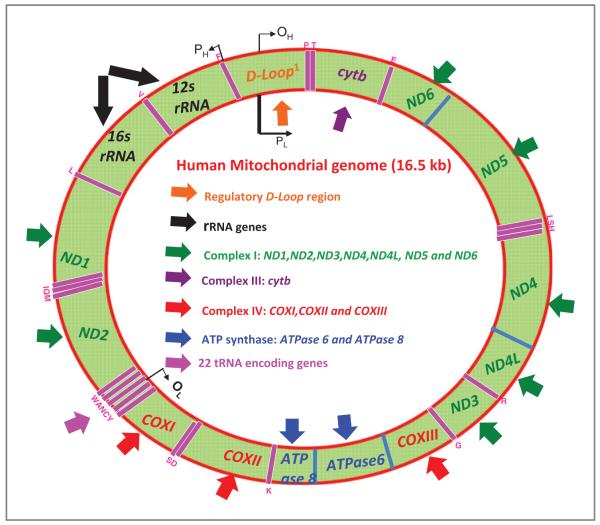
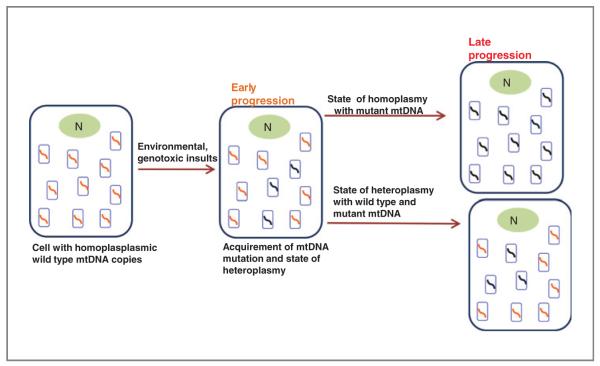
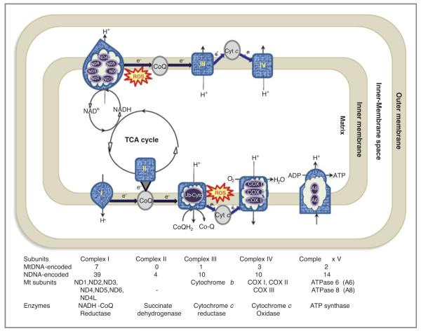
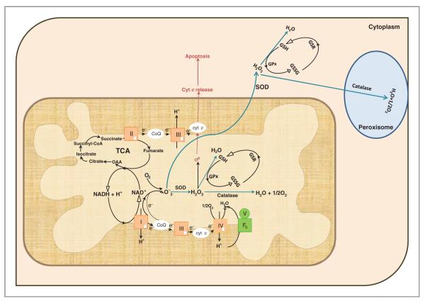
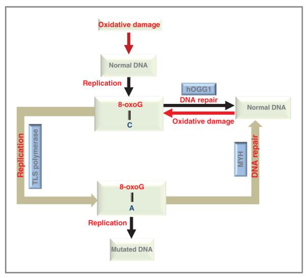
Mitochondria control essential cellular activities including generation of ATP via oxidative phosphorylation. Mitochondrial DNA (mtDNA) mutations in the regulatory D-loop region and somatic mtDNA mutations are common in primary human cancers. The biological impact of a given mutation may vary, depending on the nature of the mutation and the proportion of mutant mtDNAs carried by the cell. Identification of mtDNA mutations in precancerous lesions supports their early contribution to cell transformation and cancer progression. Introduction of mtDNA mutations in transformed cells has been associated with increased ROS production and tumor growth. Studies reveal that increased and altered mtDNA plays a role in the development of cancer but further work is required to establish the functional significance of specific mitochondrial mutations in cancer and disease progression. This review offers some insight into the extent of mtDNA mutations, their functional consequences in tumorigenesis, mitochondrial therapeutics, and future clinical application.


Figures

Figure 1
Figure 1
Organization of the human mitochondrial genome. The 16.5 kb genome encodes 37 genes including 22 for mitochondrial tRNA (20 standard amino acids, pink arrows) and 2 for the rRNA (black arrows). The rest of the 13 genes encode for subunits of the different respiratory complexes I to IV (11 genes) and ATP synthase (2 genes). Complex I (NADH dehydrogenase) is composed of 7 mitochondrial subunits (ND1, ND2, ND3, ND4, ND5, ND6,and ND4L) and 37 nuclear subunits. Complex-II (succinate dehydrogenase) is composed of 4 subunits (all being nuclear encoded) and is both a component of the ETC and an enzyme of the Krebs cycle. Complex III (cytochrome c reductase) is a complex of 11 subunits. Only subunit, cytochrome b (purple arrow), is encoded by mtDNA, the remainder 10 are nuclear encoded. Complex IV (cytochrome c oxidase) is a complex of 13 different subunits, 3 (red arrows) are encoded by mtDNA, and 10 are nuclear encoded. The ATP synthase family is composed of 14 subunits; 2 (blue arrows) are mtDNA encoded. The other 14 are nuclear encoded. The displacement loop (D-loop, orange arrows) is the main noncoding area of the mtDNA where replication occurs. The region contains promoters for the transcription of RNA from the 2 strands of mtDNA. The heavy H strand has higher guanine content, and is transcribed from the PH promoter. The light L strand is transcribed from the PL promoter. Replication of the heavy strand by DNA polymerase commences from the OH replication origin; this eventually exposes the OL origin allowing replication of the light strand.
Figure 2
Figure 2
Homoplasmic and heteroplasmic mtDNA mutations due to environmental and genotoxic damages, along with nuclear (N) genetic changes; cells may incorporate somatic mtDNA mutations and acquire a state of heteroplasmy with both wild-type and mutated mtDNA copies. Further progression of tumor cells may be aided with a homoplasmic bias with mutant mtDNA copies or an admixture of both wild-type and mutant mtDNA, a state of heteroplasmy.
Figure 3
Figure 3
Schematic representation of mitochondria complexes. The ETC in the mitochondrion is the site of oxidative phosphorylation. The NADH and succinate generated in the citric acid cycle (TCA cycle) are oxidized, providing energy to power ATP synthase. The diagram shows the complexes involved in OXPHOS (oxidative phosphorylation). Complex I (also known as NADH coQ reductase or NADH dehydrogenase) accepts electrons from the citric acid cycle (TCA cycle) electron carrier NADH, and passes them to coQ (ubiquinone; labeled CoQ), which also receives electrons from complex II, (also known as succinate dehydrogenase or succinate-Q oxidoreductase). CoQ passes electrons to complex III, (also known as Q-cytochrome c oxidoreductase or cytochrome c reductase or cytochrome b complex), which passes them to cytochrome c (Cyt c). Cyt c passes electrons to complex IV (also known as cytochrome c oxidase), which uses the electrons and hydrogen ions to reduce molecular oxygen to water. The electrochemical proton gradient allows ATP synthase (ATPase or complex V) to use the flow of H+ through the enzyme back into the matrix to generate ATP from adenosine diphosphate (ADP). The last destination for an electron along this chain is an oxygen molecule. Normally the oxygen is reduced to produce water; however, few of the electrons passing through the chain escape and oxygen is instead reduced to the superoxide radical (ROS).
Figure 4
Figure 4
Antioxidant defense mechanism in mitochondria. Mitochondria is the primary site for generation of ATP, which is the energy needed for cellular machinery. In addition to energy, ROS are produced, which results in cellular damage. The most commonly known ROS are hydrogen peroxide (H2O2), superoxide (O2), and hydroxyl ions (OH). Superoxide generated in mitochondria is converted to H2O2 by the enzyme Mn-SOD. The H2O2 is further degraded to water by 2 defense mechanisms: (i) Glutathione peroxidase (GPx) catalyzes the reaction, whereby GSH reacts with H2O2 and converts it to glutathione disulfide (GSSG) and water (H2O). Glutathione reductase (GSR) then reduces the oxidized glutathione (GSSG) to GSH. (ii) As a second mode of defense the H2O2 is converted into H2O and molecular oxygen (O2) by the mitochondrial catalase. Any superoxide that escapes mitochondria is again attacked by the SOD (Cu/Zn) present in the cytosol, which is again converted to H2O2 and this peroxide is decomposed by GPx and Catalase present in the cytoplasm and peroxisomes, respectively. The last destination for an electron along this chain is an oxygen molecule. Normally the oxygen is reduced to produce water; however, few of the electrons passing through the chain leak resulting in the generation of O2. The most common site for electron leak are complexes I and III. Superoxide is not particularly reactive by itself, but can inactivate specific enzymes or initiate the formation of OH (depicted in red in dotted lines). Accumulation of OH in mitochondria could lead to release of cyt C from mitochondria leading to apoptosis. The diagram also depicts the TCA cycle which takes place in the matrix of the mitochondria. To be noted: the diagram depicts NADH being generated by malate dehydrogenase, but in TCA cycle NADH is also generated by isocitrate dehydrogenase and α-ketoglutarate dehydrogenase.
Figure 5
Figure 5
DNA repair pathway in mitochondria. A schematic representation of BER in mitochondria. In normal DNA guanine (G) pairs with cytosine (C); G:C. In case of oxidative DNA damage, guanine is directly oxidized to 8-oxoG, which pairs against cytosine (C). The oxidatively damaged base is repaired by DNA glycosylase, here the represented glycosylase is hOGG1 (8-oxoguanine DNA glycosylase which acts both as an N-glycosylase and an AP-lyase), which specifically removes the 8-oxoG opposite C and restores the normal DNA, that is, G:C. If unrepaired, the damaged base are replicated by translesion DNA synthesis (TLS) polymerases (181), which results in an 8-oxoG opposite adenine (A). In the second defense mechanism, repair enzymes like hMYH (Mut Y homologue) act in removing adenine opposite 8-oxoG thus eliminating G:C → T:A transversions. If the 8-oxoG:A is left unrepaired and undergoes DNA replication it results in mutated DNA, that is, G:C to T:A.

Similar articles

Cited by

References

    1. Warburg O. On respiratory impairment in cancer cells. Science. 1956;124:269–70. - PubMed
    1. Warburg O. On the origin of cancer cells. Science. 1956;123:309–14. - PubMed
    1. Warburg O. Origin of cancer cells. Oncologia. 1956;9:75–83. - PubMed
    1. Attardi G, Schatz G. Biogenesis of mitochondria. Annu Rev Cell Biol. 1988;4:289–333. - PubMed
    1. Lightowlers RN, Chinnery PF, Turnbull DM, Howell N. Mammalian mitochondrial genetics: heredity, heteroplasmy and disease. Trends Genet. 1997;13:450–5. - PubMed

Publication types

Substances