. 2011 Dec 7;8(65):1748-59.
doi: 10.1098/rsif.2011.0114. Epub 2011 May 4.

Adjuvant properties of a biocompatible thermo-responsive polymer of N-isopropylacrylamide in autoimmunity and arthritis

Affiliations

Adjuvant properties of a biocompatible thermo-responsive polymer of N-isopropylacrylamide in autoimmunity and arthritis

Akhilesh Kumar Shakya et al. J R Soc Interface. .

Abstract

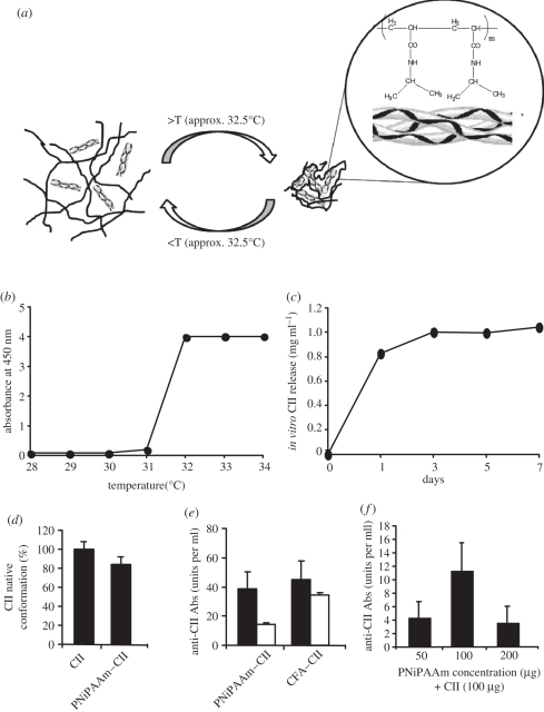
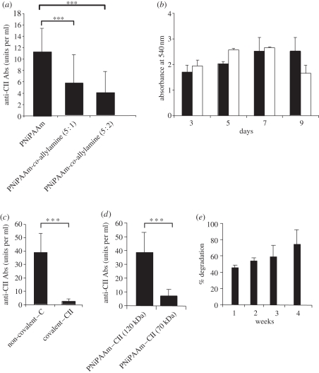
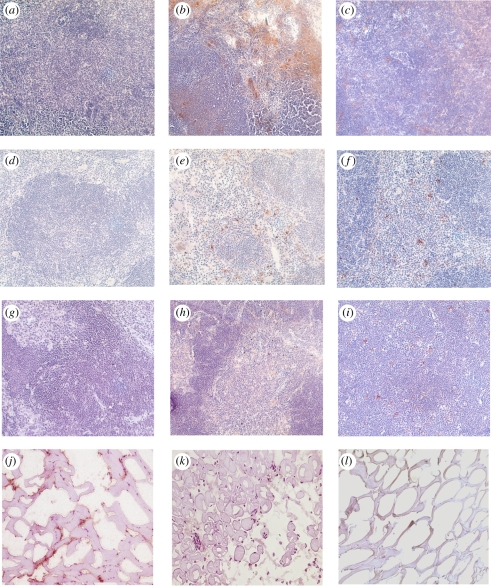
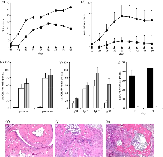
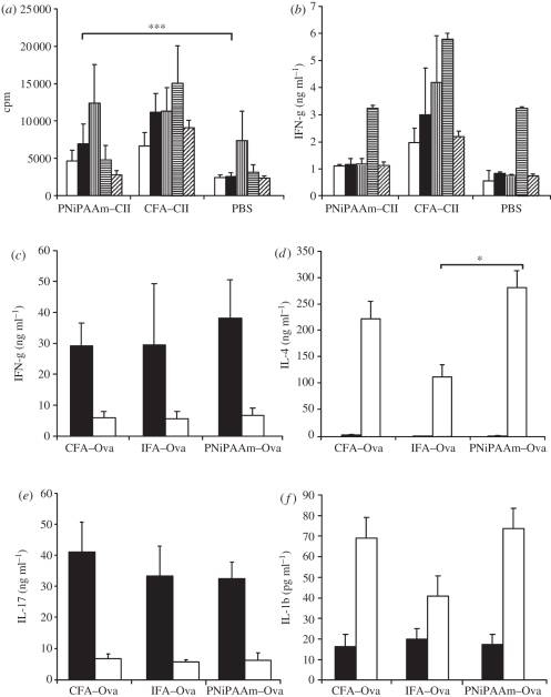
To evaluate the thermo-responsive poly(N-isopropylacrylamide) (PNiPAAm) polymer as an adjuvant, we synthesized PNiPAAm through free radical polymerization and characterized it both in vitro and in vivo. The polymer when mixed with collagen type II (CII) induced antigen-specific autoimmunity and arthritis. Mice immunized with PNiPAAm-CII developed significant levels of CII-specific IgG response comprising major IgG subclasses. Antigen-specific cellular recall response was also enhanced in these mice, while negligible level of IFN-γ was detected in splenocyte cultures, in vitro. PNiPAAm-CII-immunized arthritic mouse paws showed massive infiltration of immune cells and extensive damage to cartilage and bone. As determined by immunostaining, most of the CII protein retained its native configuration after injecting it with PNiPAAm in naive mice. Physical adsorption of CII and the high-molecular-weight form of moderately hydrophobic PNiPAAm induced a significant anti-CII antibody response. Similar to CII, mice immunized with PNiPAAm and ovalbumin (PNiPAAm-Ova) induced significant anti-ovalbumin antibody response. Comparable levels of serum IFN-γ, IL-1β and IL-17 were observed in ovalbumin-immunized mice with complete Freund, incomplete Freund (CFA and IFA) or PNiPAAm adjuvants. However, serum IL-4 levels were significantly higher in PNiPAAm-Ova and CFA-Ova groups compared with the IFA-Ova group. Thus, we show for the first time, biocompatible and biodegradable thermo-responsive PNiPAAm can be used as an adjuvant in several immunological applications as well as in better understanding of the autoimmune responses against self-proteins.


Figures

Figure 1.
Figure 1.
In vitro and in vivo characterization of PNiPAAm. Schematic of thermo-responsive behaviour of PNiPAAm with CII (a). Below LCST (approx. 32°C) (b), 100 µg of PNiPAAm mixed with 100 µg of CII (1 mg ml−1) remains in solution and above this temperature forms a lattice incorporating CII; in vitro release profile of CII from PNiPAAm at 37°C (c); percentage of released CII retaining native CII conformation from PNiPAAm–CII mixture (100 µg each at 1 : 1 ratio) in vitro (d); serum antibody response recognizing native and/or denatured conformation of CII (units per ml) (e). Optimization of PNiPAAm concentration (50–200 µg) for immunization mixed with 100 µg of CII (f) after 21 days of injection. Error bars denote ±s.e.m. (e) Black bars, native CII; white bars, denatured CII.
Figure 2.
Figure 2.
Physical and biochemical characterization of PNiPAAm. Hydrophilicity effect on the adjuvant potential of PNiPAAm (a); MTT cell proliferation assay of HT1080 cell line grown over the PNiPAAm surface (b); antibody response to CII (units per ml) in mice immunized with PNiPAAm incorporating CII in a non-covalent and covalent method (c); antibody response to CII (units per ml) in mice immunized with CII mixed with two different molecular weight forms of PNiPAAm (120 and 70 kDa) (d); gravimetric degradation kinetics of PNiPAAm at different time points in vivo (e). ***p < 0.001. Error bars denote ±s.e.m. (b) Black bars, PNiPAAm; white bars, control.
Figure 3.
Figure 3.
Bio-distribution of the biotinylated polymer in different lymphoid organs. Tissue sections from control mice (a,d,g) and mice sacrificed on day 1 (b,e,h) and day 30 (c,f,i) after biotinylated PNiPAAm-co-allylamine injection are shown. Lymph nodes (a–c), spleen (d–f) and thymus (g–i) samples were used. Liver sections did not show any staining for the polymer (data not shown). Immunostaining of CII with monoclonal antibodies recognizing native (j) CB20 or denatured (k) GB8 conformations. Control staining (l). All the images were taken at 20× magnification. (Online version in colour.)
Figure 4.
Figure 4.
PNiPAAm as an adjuvant in the induction of autoimmunity and arthritis. (a) Arthritis incidence and (b) mean arthritis score on different days are shown. Groups of 8-week-old B10.RIII mice were immunized subcutaneously at the base of the tail with 100 µg of CII emulsified in complete Freund's adjuvant (CFA–CII; n = 19), CII mixed with 100 µg of PNiPAAm (PNiPAAm–CII; n = 48) or 100 µg of ovalbumin mixed with 100 µg of PNiPAAm (PNiPAAm–Ova; n = 10) on day 0 in 200 µl volume. Mice were boosted with 50 µg of CII emulsified in incomplete Freund's adjuvant, CII mixed with PNiPAAm or ovalbumin mixed with PNiPAAm, respectively, on day 21 or 35. Results are from three experiments and all the animals were used for calculations. Sera collected on either days 21/35 (pre-boost) or days 50/60 (post-boost) were used for antibody analysis (c–e) as described in §2. CII without any adjuvant induced neither arthritis nor an anti-CII antibody response in a group of mice (n = 8). IgG subclass analysis was done in sera collected after 50 days of injection. Representative histology joints of mice (n = 3–4 in each group) from PBS (f), PNiPAAm–CII (g) and CFA–CII (h) groups. n indicates the number of mice in each group. Error bars denote ±s.e.m. All the images were taken at 20× magnification. (a,b) Solid line with oval-shaped symbols, CFA–CII; solid line with squares, PNiPAAm–CII; solid line with triangles, PNiPAAm–Ova. (c) Black bars, PNiPAAm–Ova; white bars, PNiPAAm–CII; striped bars, CFA–CII. (d) White bars, PNiPAAm–CII; striped bars, CFA–CII. (e) Black bars, PNiPAAm–Ova; white bars, PNiPAAm–CII. (Online version in colour.)
Figure 5.
Figure 5.
PNiPAAm enhances cellular activation and serum cytokine production. Groups of B10.RIII mice (n = 15) were immunized with bovine PNiPAAm–CII, CII emulsified in CFA (CFA–CII) or PBS and 10 days later splenocytes were cultured in triplicates at a concentration of 1 × 10⁶ cells per well for 72 h with medium alone, 50 µg ml−1 bovine CII, 5 µg ml−1 of concanavalin A or 0.5 µg ml−1 of anti-CD3 (clone 17A2) in DMEM + Glutamax-I supplemented with 5% heat-inactivated foetal calf serum and penicillin/streptomycin and then pulsed with [3H] thymidine for a further 15–18 h before harvesting the cells. (a) cpm denotes counts per minute. (b) Supernatant of cultured cells were collected after 72 h for IFN-γ estimation. For serum cytokine measurements, C57BL/6NJ mice (n = 15) were immunized at the base of the tail with 100 µg of ovalbumin emulsified with complete Freund's adjuvant (CFA–Ova), incomplete Freund's adjuvant (IFA–Ova) or mixed with PNiPAAm. Sera collected on days 10 and 19 were analysed for various cytokines (c–f) as described in §2. *p < 0.05; ***p < 0.001. Error bars denote ±s.e.m. n indicates the number of mice in each group. (a,b) White bars, medium; black bars, CII; bars with vertical stripes, ConA; bars with horizontal stripes, anti-CD3; bars with oblique stripes, Ova. (cf) Black bars, 10 days; white bars, 19 days.

References

    1. Monach P. A., Benoist C., Mathis D. 2004. The role of antibodies in mouse models of rheumatoid arthritis, and relevance to human disease. Adv. Immunol. 82, 217–24810.1016/S0065-2776(04)82005-4 (doi:10.1016/S0065-2776(04)82005-4) - DOI - DOI - PubMed
    1. Kannan K., Ortmann R. A., Kimpel D. 2005. Animal models of rheumatoid arthritis and their relevance to human disease. Pathophysiology 12, 167–18110.1016/j.pathophys.2005.07.011 (doi:10.1016/j.pathophys.2005.07.011) - DOI - DOI - PubMed
    1. Reed S. G., Bertholet S., Coler R. N., Friede M. 2009. New horizons in adjuvants for vaccine development. Trends Immunol. 30, 23–3210.1016/j.it.2008.09.006 (doi:10.1016/j.it.2008.09.006) - DOI - DOI - PubMed
    1. Wilson-Welder J. H., Torres M. P., Kipper M. J., Mallapragada S. K., Wannemuehler M. J., Narasimhan B. 2009. Vaccine adjuvants: current challenges and future approaches. J. Pharm. Sci. 98, 1278–131610.1002/jps.21523 (doi:10.1002/jps.21523) - DOI - DOI - PMC - PubMed
    1. Edelman R. 2002. The development and use of vaccine adjuvants. Mol. Biotechnol. 21, 129–14810.1385/MB:21:2:129 (doi:10.1385/MB:21:2:129) - DOI - DOI - PubMed

Publication types

MeSH terms