. 2011 Jul;85(14):7108-17.
doi: 10.1128/JVI.00573-11. Epub 2011 May 4.

Identification of amino acid residues important for heparan sulfate proteoglycan interaction within variable region 3 of the feline immunodeficiency virus surface glycoprotein

Affiliations

Identification of amino acid residues important for heparan sulfate proteoglycan interaction within variable region 3 of the feline immunodeficiency virus surface glycoprotein

Qiong-Ying Hu et al. J Virol. 2011 Jul.

Abstract

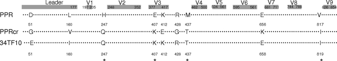
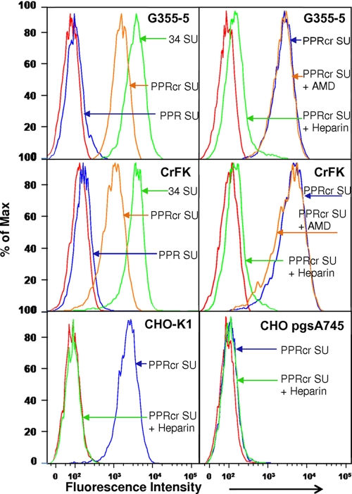
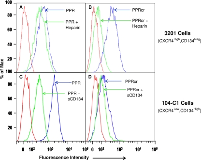
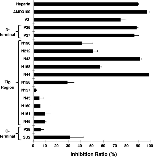
Heparan sulfate proteoglycans (HSPGs) act as binding receptors or attachment factors for the viral envelope of many viruses, including strains of HIV and feline immunodeficiency virus (FIV). The FIV gp95 glycoprotein (SU) from laboratory-adapted strains (tissue culture adapted [TCA]) such as FIV-34TF10 can bind to HSPG, whereas SU from field strains (FS) such as FIV-PPR cannot. Previous studies indicate that SU-HSPG interactions occur within the V3 loop. We utilized a series of nested V3 peptides to further map the HSPG binding sites and found that both sides of the predicted V3 loop stem were critical for the binding but not the CXCR4 binding domain near the predicted tip of the V3 loop. Neutralization assays for TCA strain entry using the same set of V3 peptides showed that peptides targeting CXCR4 or HSPG binding sites can block infection, supporting the V3 loop as a critical neutralization target. Site-directed mutagenesis identified two highly conserved arginines, R379 and R389, on the N-terminal side of the V3 stem as critical for the contact between SU and HSPG. Residues K407, K409, K410, and K412 on the C-terminal side of the V3 stem form a second nonconserved domain necessary for HSPG binding, consistent with the observed specificity distinctions with FS FIV. Our findings discriminate structural determinants important for HSPG and CXCR4 binding by FIV SU and thus further define the importance of the V3 loop for virus entry and infection.


Figures

Fig. 1.
Fig. 1.
Schematic representation of sequence alignment of PPR, PPRcr, and 34TF10 SU glycoprotein. A total of nine mutations were noted between FS FIV-PPR and TCA FIV-PPRcr. Six of the above nine amino acid positions were the same in TCA FIV-34TF10 as in FIV-PPR, and four of the six changes were common between the two TCA isolates.
Fig. 2.
Fig. 2.
PPRcr SU can bind to HSPG-expressing cells. FACS analysis of FIV SU binding to G355-5 cells (top), CrFK (middle), CHO-K1 (bottom left), and CHO-pgsA745 (bottom right). Cells were preincubated with the CXCR4 antagonist AMD3100 (0.3 μg/ml) at 4°C for 30 min prior to incubation with SU or coincubated with SU and heparin (10 μg/ml) at 4°C for 45 min. After washing, FIV SU-Fc binding was measured by using a phycoerythrin-conjugated anti-Fc antibody and then monitored by FACS analysis. Results are representative of three independent determinations.
Fig. 3.
Fig. 3.
Comparative binding of PPR and PPRcr SU to CXCR4 (3201 cells) and CD134 (104-C1 cells). (A) FS PPR SU binding to CXCR4 on 3201 cells with or without heparin. (B) TCA PPRcr SU binding to CXCR4 on 3201 cells with or without heparin. (C) FS PPR SU binding to CD134 on 104-C1 cells with or without soluble CD134 (sCD134) as competitor. (D) TCA PPRcr SU binding to CXCR4 with or without soluble CD134 as competitor. Heparin in panels A and B was incubated with SU for 45 min at 25°C, at 20 μg/ml. Soluble CD134 (25 μg/ml) in panels C and D was incubated with SU at 25°C for 30 min prior to addition to cells and then incubated for another 45 min, washed, and analyzed by FACS. Results are representative of three independent determinations.
Fig. 4.
Fig. 4.
P26 and SU2 peptides interfere with PPRcr and 34TF10 SU-Fc binding to HSPG in a dose-dependent manner. G355-5 cells were pretreated with P26 or SU2 peptide, alone or in combination, at 4°C for 30 min, and then the cells were spun down to remove the peptides and washed twice. Five hundred nanograms of PPRcr SU-Fc (A) or 34TF10 (B) was added to cells and incubated at 4°C for 45 min. After washing, SU binding was monitored by FACS analysis as described for Fig. 2. When treated separately, P26 was used at 50, 25, 10, and 5 μg/ml as final concentrations and SU2 was used at 100, 50, 25, and 10 μg/ml. When combined, P26 was used at 5 and 10 μg/ml and SU2 was used at 10 and 25 μg/ml. Percent inhibition was calculated as described in Materials and Methods. Results are means ± standard deviations for three independent determinations.
Fig. 5.
Fig. 5.
Neutralization effects of V3 peptides on 34TF10 entry into G355-5 cells. β-Gal-expressing pseudovirions with 34TF10 envelope were produced as described in Materials and Methods. All peptides were preincubated with G355-5 cells at 37°C for 60 min at a final concentration of 25 μg/ml and then cotreated with 34TF10 pseudovirions to perform a single-round infection in G355-5 cells. AMD3100 (1 μg/ml) and heparin (10 μg/ml) were used as positive controls for inhibition of entry. β-Gal assays were performed 48 h after infections. Values for inhibition by peptides, AMD3100, or heparin are percentages of the mean relative luminescence units of 34TF10 entry without any treatment, which is regarded as 100%. Results are means and standard deviations for three independent determinations.
Fig. 6.
Fig. 6.
Schematic representation of V3 in the context of SU. (A) Sequence of the V3 region, with residues relevant to HSPG binding shown in bold. Numbers above the line represent percent conservation in comparing approximately 200 V3 sequences from known FIV isolates. (B) Cartoon showing the CXCR4 and HSPG binding regions defined on FIV V3.

Similar articles

Cited by

References

    1. Akhtar J., Shukla D. 2009. Viral entry mechanisms: cellular and viral mediators of herpes simplex virus entry. FEBS J. 276:7228–7236 - PMC - PubMed
    1. Andrianov A. M., Veresov V. G. 2007. Structural analysis of the HIV-1 gp120 V3 loop: application to the HIV-Haiti isolates. J. Biomol. Struct. Dyn. 24:597–608 - PubMed
    1. Bobardt M. D., et al. 2003. Syndecan captures, protects, and transmits HIV to T lymphocytes. Immunity 18:27–39 - PubMed
    1. Cardin A. D., Weintraub H. J. 1989. Molecular modeling of protein-glycosaminoglycan interactions. Arteriosclerosis 9:21–32 - PubMed
    1. de Parseval A., et al. 2005. A highly conserved arginine in gp120 governs HIV-1 binding to both syndecans and CCR5 via sulfated motifs. J. Biol. Chem. 280:39493–39504 - PubMed

Publication types

MeSH terms

LinkOut - more resources