. 2011 Jul;85(14):6856-66.
doi: 10.1128/JVI.01843-10. Epub 2011 May 4.

Heliothis zea nudivirus 1 gene hhi1 induces apoptosis which is blocked by the Hz-iap2 gene and a noncoding gene, pag1

Affiliations

Heliothis zea nudivirus 1 gene hhi1 induces apoptosis which is blocked by the Hz-iap2 gene and a noncoding gene, pag1

Yueh-Lung Wu et al. J Virol. 2011 Jul.

Abstract

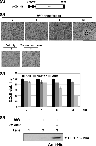
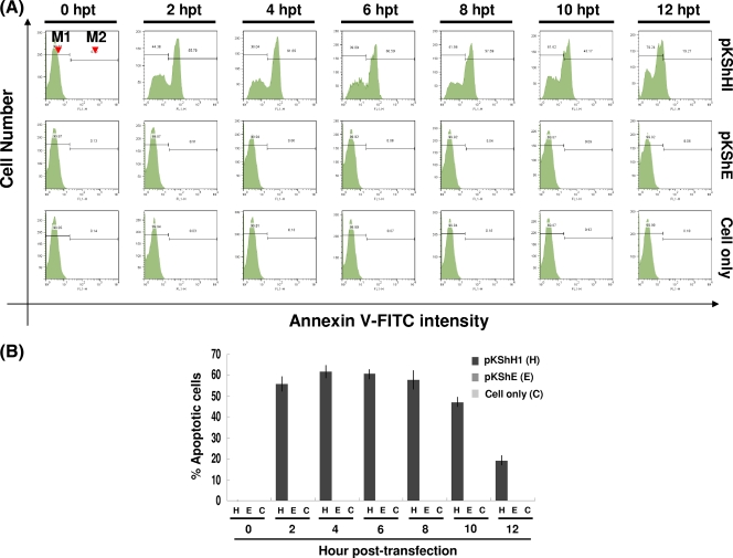
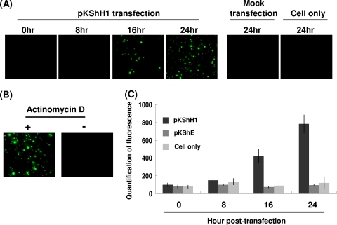
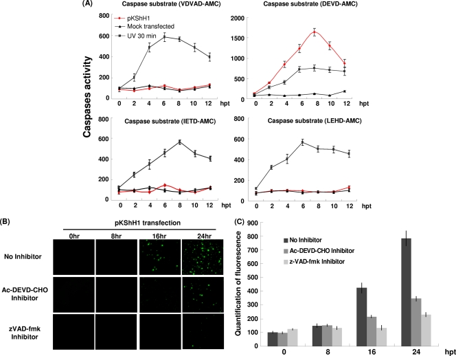
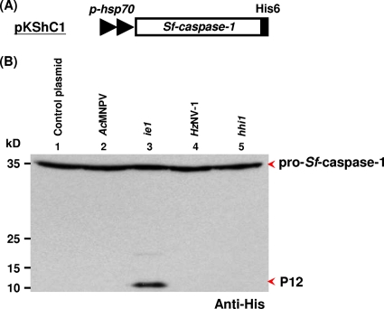
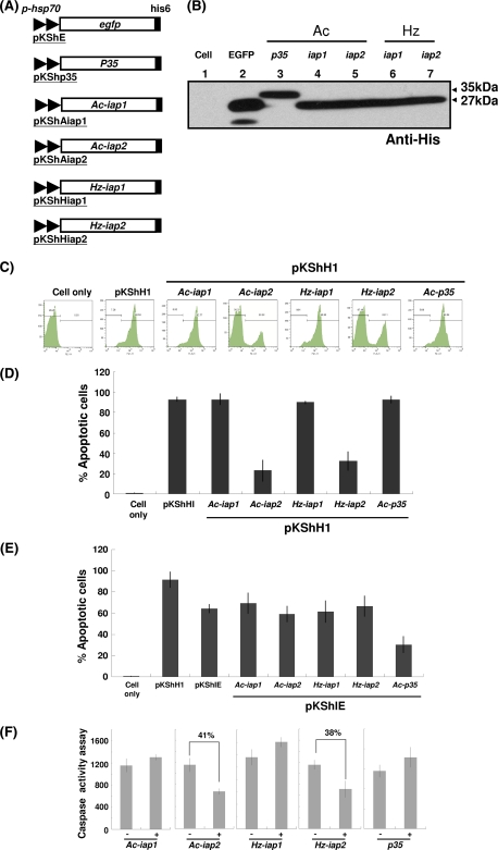
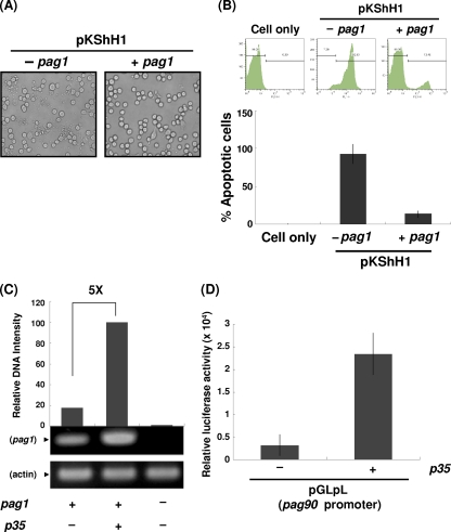
Heliothis zea nudivirus 1 (HzNV-1 or Hz-1 virus), previously regarded as a nonoccluded baculovirus, recently has been placed in the Nudivirus genus. This virus generates HzNV-1 HindIII-I 1 (hhi1) and many other transcripts during productive viral infection; during latent viral infection, however, persistency-associated gene 1 (pag1) is the only gene expressed. In this report, we used transient expression assays to show that hhi1 can trigger strong apoptosis in transfected cells, which can be blocked, at least partially, by the inhibitor of apoptosis genes Autographa californica iap2 (Ac-iap2) and H. zea iap2 (Hz-iap2). In addition to these two genes, unexpectedly, pag1, which encodes a noncoding RNA with no detectable protein product, was found to efficiently suppress hhi1-induced apoptosis. The assay of pro-Sf-caspase-1 processing by hhi1 transfection did not detect the small P12 subunit at any of the time intervals tested, suggesting that hhi1 of HzNV-1 induces apoptosis through alternative caspase pathways.


Figures

Fig. 1.
Fig. 1.
Induction of apoptosis in Sf-21 cells upon transfection with or without hhi1. (A) Schematic of the His-tagged hhi1-expression vector, pKShH1, which contains an hsp70 promoter (p-hsp) to drive hhi1 expression. (B) Monolayers of Sf-21 cells were transfected with pKShH1 by Cellfectin. Cells then were examined by light microscopy and photographed at 0, 4, 8, and 12 hpt. The arrows indicate a cluster of apoptotic bodies. Healthy and pKShE-transfected (transfection control) Sf-21 cells served as negative controls. (C) Cells were stained with calcein AM at the indicated time points, and viable cells, indicated by the exclusion of the dye, were counted under a microscope. (D) Sf-21 cells were transfected with a His-tagged hhi1 gene with or without untagged Hz-iap2 and then analyzed by Western blotting to study the expression of HHI1. Lane 1, cells only; lane 2, cells transfected with hhi1 only; lane 3, cells cotransfected with hhi1 and the antiapoptotic gene Hz-iap2.
Fig. 2.
Fig. 2.
Assessment of apoptosis resulting from hhi1 transfection in Sf-21 cells by annexin V assay. (A) Representative histograms of flow cytometry analysis showing the fluorescence intensity of annexin V-labeled apoptotic cells transfected with pKShH1 or pKShE. The annexin V-positive cells can be identified in the M2 region. M1, nonapoptotic cells; M2, apoptotic cells. (B) Bar graph showing the percentage of apoptotic cells at different time points posttransfection resulting from annexin V labeling.
Fig. 3.
Fig. 3.
Assessment of apoptosis resulting from hhi1 transfection in Sf-21 cells by TUNEL assay. (A) TUNEL assay of Sf-21 cells transfected with pKShH1 at different time points. TUNEL-positive cells were observed in Sf-21 cells transfected with pKShH1 beginning from 16 hpt. (B) Sf-21 cells left untreated or treated with the apoptosis-inducing agent actinomycin D (20 μM) were TUNEL stained at 24 h posttreatment to serve as positive and negative controls, respectively. (C) Quantification of fluorescence of TUNEL-positive cells in Sf-21 cells transfected with pKShH1 or pKShE and Sf-21 cells only at different time points.
Fig. 4.
Fig. 4.
hhi1 induces activation of an insect caspase-3-like protein. (A) Assay of the caspase activity induced by hhi1 transfection. An ApoAlert caspase assay kit (Clontech) was used to measure caspase activities in Sf-21 cells transfected with pKShH1 or pKShE (mock) at 0, 2, 4, 6, 8, 10, and 12 hpt or after being subjected to UV irradiation. The substrates VDVAD-AMC, DEVD-AMC, IETD-AMC, and LEHD-AMC were added to the cell supernatant individually. After incubation for 1 h at 37°C, enzyme activity was measured through fluorogenic cleaved substrates. (B) TUNEL assay of the hhi1-transfected cells. Sf-21 cells were transfected with pKShH1 at different time points and then incubated with or without the caspase inhibitor Ac-DEVD-CHO (10 μM) or zVAD-fmk (10 μM) before being subjected to TUNEL assay. TUNEL-positive signals were detected at 16 and 24 hpt in the absence of caspase inhibitor, but signals were clearly detected only at 24 hpt in the presence of the caspase inhibitors. (C) Bar graph representations of the TUNEL assay showing the fluorescence intensity of TUNEL-positive cells at different time points posttransfection with or without caspase inhibitor Ac-DEVD-CHO or zVAD-fmk.
Fig. 5.
Fig. 5.
hhi1 does not cause the processing of pro-Sf-caspase-1. (A) Schematic diagram of plasmid pKShC1, which expresses His-tagged Sf-caspase-1. (B) Assay monitoring the processing of Sf-caspase-1 by viral infections or plasmid transfections. Sf-21 cells first were transfected with pKShC1. At 4 hpt, cells were inoculated with AcMNPV (MOI, 1; lane 2) or HzNV-l virus (MOI, 1; lane 4). The same His-tagged Sf-caspase-1 also was cotransfected into Sf-21 cells with plasmids pKSh (control; lane 1), pKShIEΔH (expresses untagged IE1; lane 3), or pKShHIΔH (expresses untagged HHI1; lane 5) separately. Cells then were collected at 72 hpt, and both the uncleaved pro-Sf-caspase-1 and its processed P12 subunit were detected by Western blot analysis using anti-His-tagged antibody (44).
Fig. 6.
Fig. 6.
Effects of antiapoptosis genes on hhi1-induced apoptosis in Sf-21 cells. (A) Schematics of plasmids containing antiapoptosis genes. (B) Western blot analysis showing proper transient expression of antiapoptotic proteins from the plasmids shown in panel A. (C) Annexin V assay of apoptosis induction in Sf-21 cells by flow cytometry. Sf-21 cells were transfected with either hhi1 (pKShH1) alone or with anti-apoptosis gene Ac-iap1, Ac-iap2, Hz-iap1, Hz-iap2, or p35 (0.5 μg of each plasmid). Transfected cells were stained by annexin V at 2 hpt and then analyzed by flow cytometry. Apoptotic cells that stained positive for annexin V were identified in the M2 region. (D) Bar graph representation of the percentages of apoptotic cells using the data shown in panel C. (E) Apoptosis induction assay in Sf-21 cells transfected with hhi1, ie1, or hhi1 and antiapoptosis genes. (F) Caspase-specific activity assay in hhi1-transfected Sf-21 cells, which were cotransfected with or without anti-apoptosis genes, using DEVD-AMC as a substrate. Percentages of apoptosis suppression by Ac-iap2 and Hz-iap2 are indicated.
Fig. 7.
Fig. 7.
Analysis of antiapoptotic activities of pag1 in Sf-21 cells. (A) Inhibition of hhi1-induced apoptosis by pag1. Sf-21 cells (2 × 10⁵ cells per 24-well plate) were transfected with pKShH1 in the presence or absence of pKSpP1 (carrying pag1). At 12 hpt, cells were examined by light microscopy and photographed to examine the inhibition of apoptosis. (B) Flow cytometry analysis of apoptosis suppression. In this assay, apoptotic cells that stained positive by annexin V were identified in the M2 region. The bar graph below shows the percentages of apoptotic cells derived from the flow analysis. (C) PAT1 level analysis by RT-PCR. Sf-21 cells were transfected with plasmids pKSpP1 (pag1) alone or cotransfected with pKShP35 (carrying p35), and the expression of the pag1 gene product, PAT1, was analyzed by RT-PCR. The expression of actin served as a control. (D) Luciferase assay showing that p35 can activate the pag1 promoter. Plasmid pGLpL (luciferase coding region driven by the pag1 promoter) (51) was cotransfected with or without pKShP35 into Sf-21 cells. Luciferase activities from each group were measured at 48 hpt.

Similar articles

Cited by

References

    1. Ahmad M., et al. 1997. Spodoptera frugiperda caspase-1, a novel insect death protease that cleaves the nuclear immunophilin FKBP46, is the target of the baculovirus antiapoptotic protein p35. J. Biol. Chem. 272:1421–1424 - PubMed
    1. Alimonti J. B., Ball T. B., Fowke K. R. 2003. Mechanisms of CD4+ T lymphocyte cell death in human immunodeficiency virus infection and AIDS. J. Gen. Virol. 84:1649–1661 - PubMed
    1. Ayres M. D., Howard S. C., Kuzio J., Lopez-Ferber M., Possee R. D. 1994. The complete DNA sequence of Autographa californica nuclear polyhedrosis virus. Virology 202:586–605 - PubMed
    1. Burand J. P., Stiles B., Wood H. A. 1983. Structural and intracellular proteins of the nonoccluded baculovirus HZ-1. J. Virol. 46:137–142 - PMC - PubMed
    1. Chao Y. C., et al. 1998. A 2.9-kilobase noncoding nuclear RNA functions in the establishment of persistent Hz-1 viral infection. J. Virol. 72:2233–2245 - PMC - PubMed

Publication types

Substances

LinkOut - more resources