. 2011 Jul;85(14):6867-81.
doi: 10.1128/JVI.00229-11. Epub 2011 May 4.

HIV-1 Nef disrupts intracellular trafficking of major histocompatibility complex class I, CD4, CD8, and CD28 by distinct pathways that share common elements

Affiliations

HIV-1 Nef disrupts intracellular trafficking of major histocompatibility complex class I, CD4, CD8, and CD28 by distinct pathways that share common elements

Jolie A Leonard et al. J Virol. 2011 Jul.

Abstract

The Nef protein is an important HIV virulence factor that promotes the degradation of host proteins to augment virus production and facilitate immune evasion. The best-characterized targets of Nef are major histocompatibility complex class I (MHC-I) and CD4, but Nef also has been reported to target several other proteins, including CD8β, CD28, CD80, CD86, and CD1d. To compare and contrast the effects of Nef on each protein, we constructed a panel of chimeric proteins in which the extracellular and transmembrane regions of the MHC-I allele HLA-A2 were fused to the cytoplasmic tails of CD4, CD28, CD8β, CD80, CD86, and CD1d. We found that Nef coprecipitated with and disrupted the expression of molecules with cytoplasmic tails from MHC-I HLA-A2, CD4, CD8β, and CD28, but Nef did not bind to or alter the expression of molecules with cytoplasmic tails from CD80, CD86, and CD1d. In addition, we used short interfering RNA (siRNA) knockdown and coprecipitation experiments to implicate AP-1 as a cellular cofactor for Nef in the downmodulation of both CD28 and CD8β. The interaction with AP-1 required for CD28 and CD8β differed from the AP-1 interaction required for MHC-I downmodulation in that it was mediated through the dileucine motif within Nef (LL(164,165)AA) and did not require the tyrosine binding pocket of the AP-1 μ subunit. In addition, we demonstrate a requirement for β-COP as a cellular cofactor for Nef that was necessary for the degradation of targeted molecules HLA-A2, CD4, and CD8. These studies provide important new information on the similarities and differences with which Nef affects intracellular trafficking and help focus future research on the best potential pharmaceutical targets.


Figures

Fig. 1.
Fig. 1.
Nef downmodulates HLA-A2, A2/CD4, A2/CD8β, and A2/CD28 in CEM T cells. (A) Diagram of chimeric molecules. (B) Flow-cytometric analysis of CEM-SS T cells expressing the indicated chimeric proteins and transduced with the indicated adenoviral vector. Cells were analyzed at 3 days postinfection (dpi). (C) Quantitation of flow cytometry experiments shown in panel B. The mean fold downmodulation ± standard deviations is shown. n=3. (D) Flow-cytometric analysis of CEM-SS T cells transduced with a retroviral vector expressing GFP alone or NL43 Nef and the GFP reporter. (E) Quantitation of flow cytometry experiments shown in panel D. The mean fold downmodulation of GFP+ cells ± standard deviations is shown. n=3. (F) Flow-cytometric analysis of SupT1 cells transduced with the indicated adenoviral vector. Cells were analyzed at 3 dpi. Sup T1 cells express endogenous CD4, CD28, and CD8.
Fig. 2.
Fig. 2.
NL43 Nef does not downmodulate CD80, CD86, or CD1d. (A) Flow-cytometric analysis of U937 cells and THP-1 cells transduced with the indicated retroviral vector and treated as indicated with LPS, PMA, or DMSO (Undiff.). The cells were analyzed 5 days postinfection (dpi). (B) Quantitation of flow cytometry experiments shown in panel A. The mean fold downmodulation ± standard deviations is shown. n=3. (C) Flow-cytometric analysis of THP-1 cells treated as described for panel A. The surface expression levels of endogenous HLA-A2, CD4, and CD1d expression were measured at 5 dpi. (D) Quantitation of flow cytometry experiments shown in panel C. The mean fold downmodulation ± standard deviation is shown. n=3. (E) Flow-cytometric analysis of primary antigen-presenting cells that were transduced with the indicated adenoviral vector. Endogenous HLA-A2, CD4, CD80, and CD86 expression levels were assessed at 3 dpi. Primary macrophages were GM-CSF treated prior to transduction; dendritic cells were GM-CSF, IL-4, and TNF-α treated prior to transduction. (F) Quantitation of flow cytometry experiments shown in panel E. The mean fold downmodulation ± standard deviations is shown. n=3.
Fig. 3.
Fig. 3.
Nef-induced downmodulation of MHC-I, CD4, CD8β, and CD28 is conserved across multiple clades. (A) Quantitation of downmodulation of the indicated molecule in CEM-SS cells following transduction with bicistronic murine retroviral vectors expressing the indicated Nef. The mean fold downmodulation of each molecule in GFP-positive cells as determined by flow cytometry is shown. n=3. The inset displays the data for CD28, CD1d, CD80, and CD86, with an expanded y axis to highlight differences among these molecules that are of a small magnitude. The statistical significance was determined by two-way analysis of variance. (B) Quantitation of Nef-dependent downmodulation of the indicated molecule by each Nef variant. The mean fold downmodulation in the GFP-positive cells ± standard deviations is shown. n=3. (C) Relative ability of Nef variants to downmodulate each target protein, plotted as fold downmodulation. YU-2 was removed as an outlier.
Fig. 4.
Fig. 4.
CD4 and CD8 are downmodulated by Nef in HIV-1-infected PBMCs. Flow-cytometric analysis of PBMCs infected with HXB ePLAP HIV with or without Nef, pseudotyped with the HXB envelope, is shown. Surface marker and intracellular Gag stains were performed at 3 days postinfection (dpi).
Fig. 5.
Fig. 5.
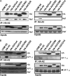
Nef physically associates with the cytoplasmic domains of HLA-A2, CD4, CD8β, and CD28 and recruits AP-1 to HLA-A2, CD8β, and CD28. Immunoprecipitation (IP) and Western blot analysis of the indicated chimeric molecule expressed in CEM T cells transduced with the indicated adenoviral vector are shown. The cells were harvested at 3 days postinfection (dpi) after an overnight incubation in ammonium chloride to inhibit degradation. Lysates were immunoprecipitated with an antibody directed against HLA-A2 (BB7.2), and the presence of HA-HLA-A2, Nef (A), or AP-1 (B) was detected by Western blot analysis.
Fig. 6.
Fig. 6.
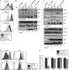
Nef recruits AP-1 for the downmodulation of CD8β and CD28 in a dileucine-dependent manner. (A) Flow-cytometric analysis of CEM SS T cells expressing the indicated chimeric protein and transduced with the indicated adenoviral vector. Nef LL164,165AA is indicated as xLL. (B) Immunoprecipitation and Western blot analysis of the indicated chimeric molecule expressed in CEM T cells transduced with the indicated adenoviral vector. The cells were harvested at 3 days postinfection (dpi). Lysates were immunoprecipitated with an antibody directed against HLA-A2 (BB7.2), and the presence of Nef, AP-1 subunits, and MHC-I (HA) were detected by Western blot analysis. Intervening lanes were removed, as indicated by the white gap. The data are representative of four independent experiments. (C) Immunoprecipitation and Western blot analysis of the indicated chimeric molecule expressed in CEM T cells transduced with the indicated adenoviral vector and treated as described for panel B. AP-2 coprecipitation is representative of two of four experiments. (D) Flow-cytometric analysis of SupT1 T cells transduced with an adenoviral vector expressing Nef and a retroviral vector expressing the indicated μ subunit of AP-1. TBPM, tyrosine binding pocket mutant μ subunit of AP-1; Wt μ, wild-type μ subunit of AP-1. The cells were analyzed by flow cytometry at 3 dpi. (E) Quantitation of flow data shown in panel D. The relative fold downmodulation, where the fold downmodulation of each molecule in the presence of Wt AP-1 is set to 100%, is shown. Error bars represent standard deviations; ***, P < 0.00001; n=3.
Fig. 7.
Fig. 7.
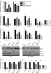
Nef requires AP-1 and β-COP for maximal downmodulation of CD8 and CD28. (A) Flow-cytometric analysis of CEM SS cells expressing the indicated chimeric protein and transduced with lentivirus expressing shRNA and GFP and subsequently transduced with the indicated adenoviral vector at 3 days after lentivirus infection. Flow-cytometric analysis was performed at 3 days after adenoviral transduction. Histograms represent GFP-expressing cells. (B) Quantitation of the flow data shown in panel A. The fold downmodulation of the indicated molecule in the presence of shRNA is shown. (C) Western blot analysis confirming the specific knockdown of AP-1 subunits in CEM SS cells. (D) Western blot analysis confirming adaptor protein knockdown in SupT1 cells. (E) Relative downmodulation of the indicated molecule in SupT1 T cells transduced with the indicated lentivirus expressing shRNA and GFP plus a bicistronic retrovirus expressing Nef and a PLAP marker gene. shNC, negative control. The cells were analyzed by flow cytometry at 3 days after retroviral transduction. The fold downmodulation normalized to the negative control in cells positive for both GFP and PLAP is shown. *, P < 0.05; **, P < 0.01; ***, P < 0.001.
Fig. 8.
Fig. 8.
CD28 is recycled rapidly in Nef-expressing cells. The measurement of recycling in CEM T cells expressing HLA-A2 or HLA-A2/CD28 and transduced with the indicated adenovirus is shown. Cells were harvested at 3 days postinfection (dpi), incubated with 150 mg/ml cycloheximide for 2 h to inhibit protein synthesis, and stripped of stainable HLA-A2 by removing the β2-microglobulin with an acid wash (50 mM glycine, 100 mM NaCl, pH 3.4). The cells then were incubated at 37°C in culture medium plus cycloheximide. Triplicate samples were removed to ice at the indicated time points, stained for surface HLA-A2 expression (BB7.2), and analyzed by flow cytometry. Recycling is plotted as a percentage of steady-state levels, where the mean fluorescence of each time point was divided by the mean fluorescence of cells that were not acid stripped. n = 2.
Fig. 9.
Fig. 9.
β-COP is required for Nef-mediated degradation of HLA-A2/CD8β. (A) Quantitation of relative downmodulation (normalized to the control) of the indicated molecule expressed in CEM-SS cells transduced with the indicated lentivirus expressing shRNA and a retroviral vector expressing Nef. The cells were harvested at 3 days after retroviral transduction. n=3. (B and C) Quantitation of the fold downmodulation of the indicated molecule expressed in CEM-SS cells (B) or Sup T1 cells (C) transduced with a retroviral vector expressing Nef or the indicated Nef mutants. Cells with equal GFP expression, to control for Nef levels, were analyzed at 3 days postinfection (dpi). The mean fold downmodulation ± standard deviations is shown. n=3. *, P < 0.05; **, P < 0.01; ***, P < 0.001. (D) Western blot analysis of endonuclease H (endo H)-treated lysates from the cells described in panel A. (E) Quantitation of endo H-resistant bands. The endo H-resistant bands (indicated by “R” in the figure) were quantified and normalized to the loading control, and shNC was set to 100% remaining. *, P < 0.05; **, P < 0.01; ***, P < 0.001; n=3.
Fig. 10.
Fig. 10.
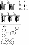
(A and B) Schematic representation of the two AP-1 binding sites that Nef utilizes. (A) When bound to the MHC-I cytoplasmic domain, Nef recruits AP-1 through the tyrosine recognition site in the AP-1μ subunit (91). In this model, the three-way complex requires amino acid residues in both MHC-I, especially Y320, as well as in Nef, M20 in particular (although assays using purified proteins and a Nef-MHC-I cytoplasmic tail fusion protein did not demonstrate a requirement for M20) (62, 72, 79, 91). When bound to the CD8 or CD28 cytoplasmic tail, Nef recruits AP-1 through the dileucine recognition site at the interface between the γ and σ subunits of AP-1 (47). (C to E) Schematic representation of the Nef domains involved in β-COP recruitment. (C) Nef residues R17 and R19 are required for β-COP involvement in MHC-I degradation (77). (D) A diacidic motif in Nef, EE155,156, is required for β-COP involvement in CD4 degradation (29, 68, 77). (E) Either or both Nef domains likely participate in β-COP recruitment for the degradation of CD8 by Nef. (F) Model of the trafficking pathways Nef uses to achieve the downmodulation and degradation of target proteins. Nef blocks anterograde transport in an AP-1-dependent pathway (46, 72, 83). Nef also induces the internalization of target molecules at the plasma membrane through an association with AP-1 or AP-2 (11, 16, 24, 44, 59, 77, 80, 84). These pathways likely converge at a β-COP-dependent step (77). Because AP-1 knockdown did not block the β-COP-dependent degradation of A2/CD8, Nef may be able to direct some targets into endolysosomal compartments by a pathway in which only β-COP recruitment is necessary and clathrin-associated adaptor proteins are dispensable. PM stands for plasma membrane, LY indicates lysosome, and LE/MVB indicates late endosome or multivesicular body compartments.

Similar articles

Cited by

References

    1. Adachi A., et al. 1986. Production of acquired immunodeficiency syndrome-associated retrovirus in human and nonhuman cells transfected with an infectious molecular clone. J. Virol. 59:284– 291 - PMC - PubMed
    1. Allan V. J., Kreis T. E. 1986. A microtubule-binding protein associated with membranes of the Golgi apparatus. J. Cell Biol. 103:2229–2239 - PMC - PubMed
    1. Arold S. T., Baur A. S. 2001. Dynamic Nef and Nef dynamics: how structure could explain the complex activities of this small HIV protein. Trends Biochem. Sci. 26:356–363 - PubMed
    1. Benichou S., et al. 1994. Physical interaction of the HIV-1 Nef protein wih β-cop, a component of non-clathrin coated vesicles essential for membrane traffic. J. Biol. Chem. 269:30073–30076 - PubMed
    1. Benson R. E., Sanfridson A., Ottinger J. S., Doyle C., Cullen B. R. 1993. Downregulation of cell-surface CD4 expression by simian immunodeficiency virus Nef prevents viral super infection. J. Exp. Med. 177:1561–1566 - PMC - PubMed

Publication types