. 2011 Jul;85(14):6893-905.
doi: 10.1128/JVI.00274-11. Epub 2011 May 4.

Identification of a novel neuropathogenic Theiler's murine encephalomyelitis virus

Affiliations

Identification of a novel neuropathogenic Theiler's murine encephalomyelitis virus

Matthew R Buckwalter et al. J Virol. 2011 Jul.

Abstract

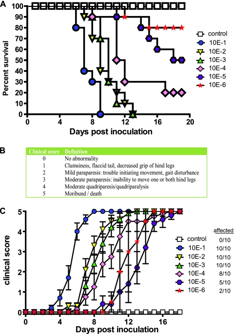
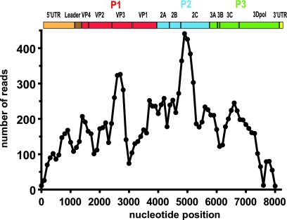
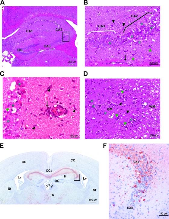
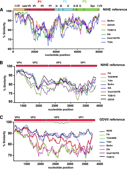
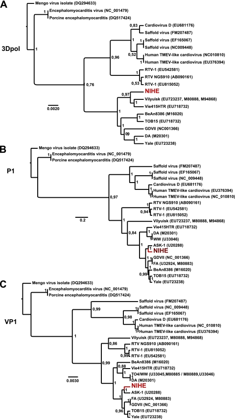
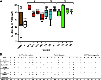
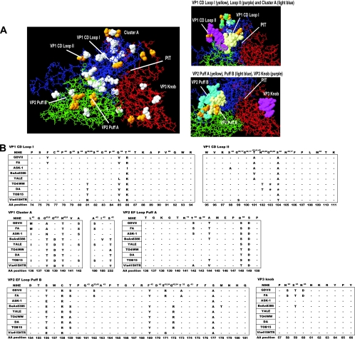
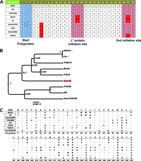
Theiler's murine encephalitis viruses (TMEV) are divided into two subgroups based on their neurovirulence. Persistent strains resemble Theiler's original viruses (referred to as the TO subgroup), which largely induce a subclinical polioencephalomyelitis during the acute phase of the disease and can persist in the spinal cord of susceptible animals, inducing a chronic demyelinating disease. In contrast, members of the neurovirulent subgroup cause an acute encephalitis characterized by the rapid onset of paralysis and death within days following intracranial inoculation. We report herein the characterization of a novel neurovirulent strain of TMEV, identified using pyrosequencing technology and referred to as NIHE. Complete coverage of the NIHE viral genome was obtained, and it shares <90% nucleotide sequence identity to known TMEV strains irrespective of subgroup, with the greatest sequence variability being observed in genes encoding the leader and capsid proteins. The histopathological analysis of infected brain and spinal cord demonstrate inflammatory lesions and neuronal necrosis during acute infection with no evidence of viral persistence or chronic disease. Intriguingly, genetic analysis indicates the putative expression of the L protein, considered a hallmark of strains within the persistent subgroup. Thus, the identification and characterization of a novel neurovirulent TMEV strain sharing features previously associated with both subgroups will lead to a deeper understanding of the evolution of TMEV strains and new insights into the determinants of neurovirulence.


Figures

Fig. 1.
Fig. 1.
Survival and kinetics of disease progression for animals inoculated with NIHE. (A) Five-week-old C57BL/6 mice were inoculated IC with 10× dilutions (10−1 to 10−6) of infected mouse brain homogenates (n = 10 per group); control mice (white boxes) received 10−1 brain homogenate derived from noninfected animals. Animals were monitored daily, and Kaplan-Meier curves illustrate the kinetics of survival. (B) Clinical scoring system used to evaluate disease progression in mice following IC inoculation with NIHE. (C) Groups of animals were monitored daily for the presence of disease. Clinical scores for symptomatic animals within each group were averaged. The number of infected animals per group is noted in the figure key.
Fig. 2.
Fig. 2.
Complete and redundant coverage of the NIHE genome was obtained by pyrosequencing. Individual sequencing reads (average length of 330 bp) were assembled de novo, and the number of sequencing reads was determined for each 100-bp segment along the length of the genome. An organizational map of the TMEV genome is aligned at the top of the figure. Gene regions are drawn to scale, and P1 (red), P2 (blue), and P3 (green) are highlighted.
Fig. 3.
Fig. 3.
Histopathological features and viral tropism within the brain and spinal cord of C57BL/6 mice 8 days after the intracranial inoculation of NIHE virus. (A to D) HE staining. Severe acute necrotizing polioencephalitis and poliomyelitis characterized by perivascular cuffs (arrows), neuronal necrosis (blue stars), satellitosis and neuronophagia (green triangles), and gliosis (black arrowheads). (A) Hippocampus (original magnification, ×100). (B) Hippocampus (Ammon's horn) (original magnification, ×200). (C) Hippocampus (original magnification, ×400). (D) Spinal cord (ventral horns) (original magnification, ×200). GM, gray matter of the spinal cord; WM, white matter of the spinal cord. (E and F) Immunostaining against Theiler's virus (strain DA). (E) Many viral neuronal bodies and processes are labeled in the Ammon's horn of the hippocampus and in thalamic nuclei. Original magnification, ×20. (F) In the Ammon's horn, many virus-positive neurons are seen and are necrotic in CA1 and CA2; in CA3 only a few neurons are infected and necrotic. Original magnification, ×200. CA, Ammon's horn; DG, dentate gyrus; CC, cerebral cortex; Cca, corpus callosum; Lv, lateral ventricle; St, striatum; Th, thalamus; CA1, CA2, and CA3, fields of the Ammon's horn; 3rdV, third ventricle.
Fig. 4.
Fig. 4.
Sequence similarity for the complete genome and P1 regions of TMEV strains compared to those of NIHE and GDVII. (A) Sequence identity for all five TMEV strains with complete genome sequence information was obtained by comparing 20-bp segments along their entire genome to the sequence of NIHE. (B) Sequence identity of full-length and partially sequenced TMEV strains using NIHE as a reference. (C) Sequence identity of all TMEV stains (including NIHE) compared to the GDVII strain as the reference sequence. All sliding-window graphs were generated using Simplot 3.2 software as described in Materials and Methods. The x axis for all graphs is the nucleotide position, and the y axis is the percent similarity. A genetic map of TMEV is shown at the top of each graph, illustrating the organization and relative size of the individual genomic regions.
Fig. 5.
Fig. 5.
Phylogenetic relationships between NIHE and select cardioviruses based on the nucleotide sequences of 3Dpol, P1, and VP1 regions. Sequences were aligned and phylogenetic analysis was performed as outlined in Materials and Methods. Phylogenetic relationships are shown for sequences of the 3Dpol (A), P1 (B), and VP1 (C) regions of the genome. Numbers at the nodes represent posterior probabilities, and the genetic distance is indicated by the bar at the bottom of each panel. The position of the NIHE strain is marked in red.
Fig. 6.
Fig. 6.
Percent identity for individual proteins of TMEV strains compared to NIHE proteins. Amino acid sequences for all available TMEV strains were compared to the NIHE sequence for each gene product. (A) The percent identity was determined, and whisker plots were generated showing the mean, minimum, and maximum values. Numbers above each plot identify how many amino acids are within each protein. Proteins are presented according to genome structure, and P1 (red), P2 (blue), and P3 (green) regions are colored according to the description in previous figure legends. (B) Amino acid alignments for eight TMEV strains are compared to the sequence of NIHE. The amino acid sequence for the Ser/Thr-rich region, Theilio domain, and L/VP4 cleavage sites are shown for the NIHE L protein along the top row. Dashes indicate identity to the NIHE sequence, and the single-letter codes for nonidentical amino acids are shown. Amino acid positions that have been implicated in a particular phenotype or function are annotated with a superscript number indicating the respective citation.
Fig. 7.
Fig. 7.
Analysis of the NIHE capsid structure formed by VP1, VP2, and VP3 proteins. Capsid proteins (VP1, VP2, and VP3) were aligned and overlaid onto the three-dimensional structure of the BeAn8386 strain using DeepView/Swiss-pdbViewer (version 4.0.1). (A) Quaternary structure with the individual proteins VP1 (blue), VP2 (green), and VP3 (red) indicated. White space-filled molecules indicate amino acid positions that differ between the sequence of BeAn8386 and NIHE. Amino acid positions that are unique to NIHE (compared to all TMEV strains) are highlighted in yellow. Highly variable regions, loops, puffs, and a canyon or “pit” are annotated. Also shown, to the right, are a close-up of VP1 CD loops I and II and the cluster A region (upper), as well as VP2 puff A and B and the VP3 knob (lower). (B) Amino acid alignments for the indicated region of all TMEV strains compared to the NIHE sequence. Dashes indicate identity to the NIHE sequence, and the single-letter codes for nonidentical amino acids are shown. Amino acid positions and annotations are the same as those described in the legend to Fig. 6.
Fig. 8.
Fig. 8.
Alternative AUG initiation sequence present for the L* gene within the NIHE genome. Nucleotide sequences were aligned for TMEV strains and compared to the sequence of NIHE. (A) The canonical start sequence (AUG) is outlined in blue, and both downstream initiating codon sequences are outlined in red. Sequences not identical to that of NIHE are highlighted in red. (B) Nucleotide sequences potentially coding for L* were aligned, and a midrooted tree was constructed using the maximum-likelihood method. There were a total of 156 positions in the final data set. Numbers at the nodes represent posterior probabilities, and genetic distance is indicated by the bar at the bottom of the panel. GDVII and FA strains were included in the analysis, although they do not efficiently produce L* (indicated by an asterisk). (C) Nucleotide sequences were aligned for all TMEV strains with available sequence information and compared to the sequence of NIHE. Only amino acid positions with discrepancies between strains are shown. The NIHE sequence is presented along the top row. Dashes indicate identity to the NIHE sequence, and the single-letter codes for nonidentical amino acids are shown.

Similar articles

Cited by

References

    1. Aubert C., Brahic M. 1995. Early infection of the central nervous system by the GDVII and DA strains of Theiler's virus. J. Virol. 69:3197–3200 - PMC - PubMed
    1. Aubert C., Chamorro M., Brahic M. 1987. Identification of Theiler's virus infected cells in the central nervous system of the mouse during demyelinating disease. Microb. Pathog. 3:319–326 - PubMed
    1. Bandyopadhyay P. K., Pritchard A., Jensen K., Lipton H. L. 1993. A three-nucleotide insertion in the H stem-loop of the 5′ untranslated region of Theiler's virus attenuates neurovirulence. J. Virol. 67:3691–3695 - PMC - PubMed
    1. Bureau J. F., et al. 1992. The interaction of two groups of murine genes determines the persistence of Theiler's virus in the central nervous system. J. Virol. 66:4698–4704 - PMC - PubMed
    1. Camacho C., et al. 2009. BLAST+: architecture and applications. BMC Bioinformatics 10:421. - PMC - PubMed

Publication types

LinkOut - more resources