. 2011 Jul;85(14):7353-62.
doi: 10.1128/JVI.00141-11. Epub 2011 May 4.

Sprouty 2 binds ESCRT-II factor Eap20 and facilitates HIV-1 gag release

Affiliations

Sprouty 2 binds ESCRT-II factor Eap20 and facilitates HIV-1 gag release

G N Medina et al. J Virol. 2011 Jul.

Abstract

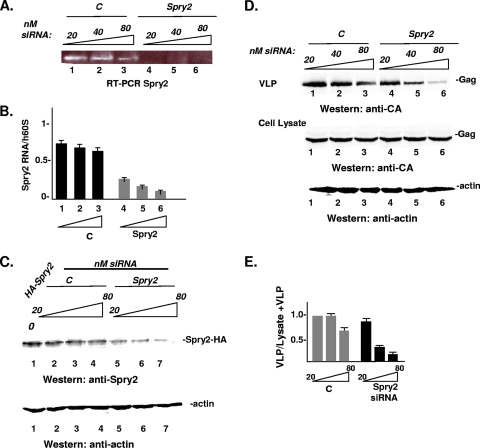
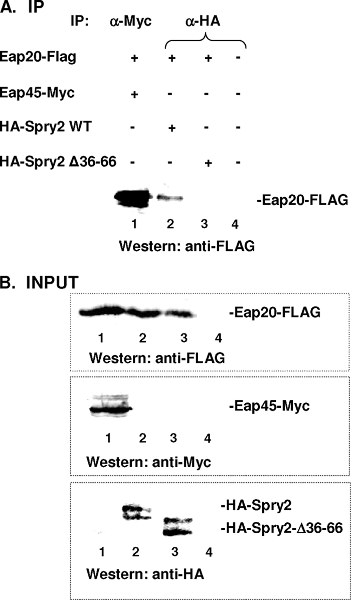
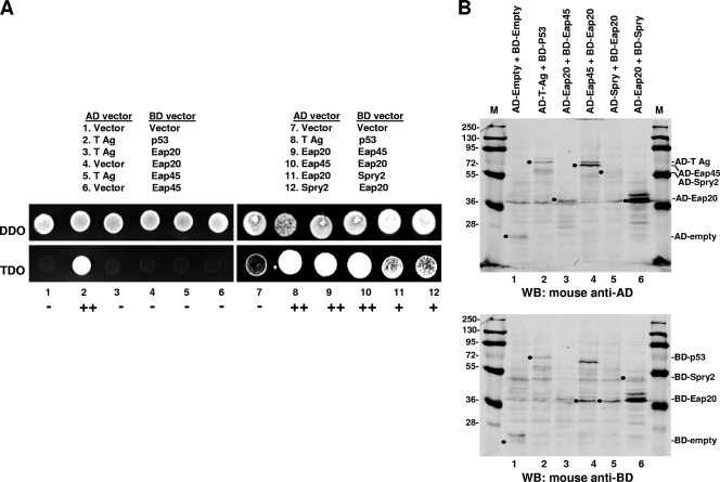
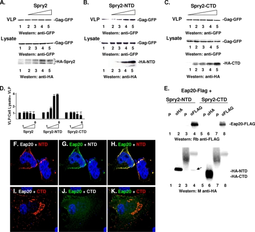
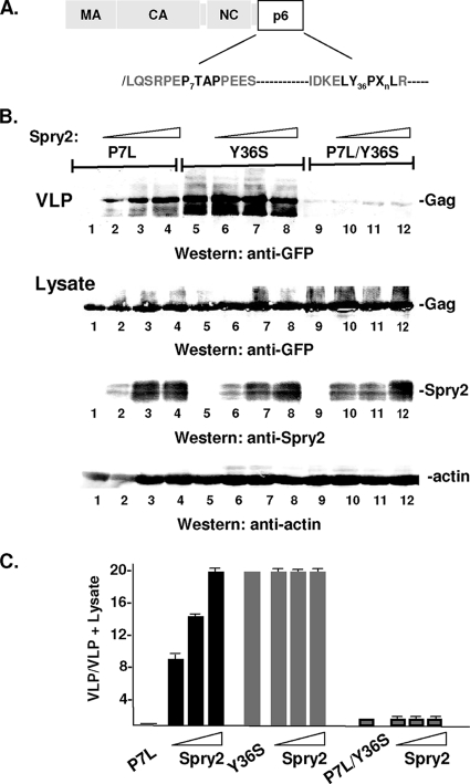
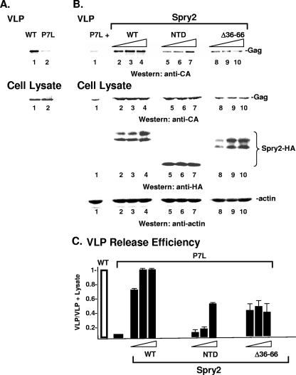
The four ESCRT (endocytic sorting complexes required for transport) complexes (ESCRT-0, -I, -II, and -III) normally operate sequentially in the trafficking of cellular cargo. HIV-1 Gag trafficking and release as virus-like particles (VLPs) require the participation of ESCRTs; however, its use of ESCRTs is selective and nonsequential. Specifically, Gag trafficking to release sites on the plasma membrane does not require ESCRT-0 or -II. It is known that a bypass of ESCRT-0 is achieved by the direct linkage of the ESCRT-I component, Tsg101, to the primary L domain motif (PTAP) in Gag and that bypass of ESCRT-II is achieved by the linkage of Gag to ESCRT-III through the adaptor protein Alix. However, the mechanism by which Gag suppresses the interaction of bound ESCRT-I with ESCRT-II is unknown. Here we show (i) that VLP release requires the steady-state level of Sprouty 2 (Spry2) in COS-1 cells, (ii) that Spry2 binds the ESCRT-II component Eap20, (iii) that binding Eap20 permits Spry2 to disrupt ESCRT-I interaction with ESCRT-II, and (iv) that coexpression of Gag with a Spry2 fragment that binds Eap20 increases VLP release. Spry2 also facilitated release of P7L-Gag (i.e., release in the absence of Tsg101 binding). In this case, rescue required the secondary L domain (YPX(n)L) in HIV-1 Gag that binds Alix and the region in Spry2 that binds Eap20. The results identify Spry2 as a novel cellular factor that facilitates release driven by the primary and secondary HIV-1 Gag L domains.


Figures

Fig. 1.
Fig. 1.
Depletion of the endogenous pool of Spry2 inhibits HIV-1 Gag release. (A) Qualitative analysis of Spry2 mRNA expression. COS-1 cells were treated with siRNA against Spry2 or against a nonspecific target (C) 24 h prior to transfection with DNA encoding Gag-GFP. A total of 48 h post-DNA transfection, total RNA was harvested, and cDNAs were generated. The blot shows Spry2 mRNA expression following treatment of COS-1 cells with control or targeted siRNA. (B) Analysis of Spry2 depletion by qPCR. cDNA levels were quantified by qPCR analysis using primers specific to Spry2. The data were normalized to the housekeeping gene h60S levels. The results represent the average of three independent experiments. (C) Analysis of Spry2 depletion by Western analysis. Cell lysates prepared as described in Materials and Methods were centrifuged to remove particulate material and then analyzed by Western blotting for exogenous Spry2 protein expression using antibody directed to the Spry2 C-terminal region. (D) Effect of Spry2 depletion on VLP release from cells. Gag proteins in cell lysates and VLPs in media from samples treated with control (lanes 1 to 3) or targeted siRNA (lanes 4 to 6) were isolated as described in Materials and Methods and analyzed by SDS-PAGE and Western blotting. (E) Semiquantitative analysis of VLP release. The panel shows the ratio of the Gag signal in VLP isolated from the media to the sum of the Gag signal in VLPs plus the cell lysate.
Fig. 2.
Fig. 2.
HA-Spry2 coimmunoprecipitates with Eap20-FLAG. (A) Immunoprecipitation reactions. DNA encoding Myc-tagged Eap45 (lane 1), wild-type (WT) HA-tagged Spry2 (lane 2), or HA-Spry2 Δ36-66 (lane 3) was coexpressed with FLAG-tagged Eap20. Eap20-FLAG was immunoprecipitated (IP) from the lysates with an anti (α)-Myc (lane 1) or anti-HA antiserum (lanes 2 and 3) and detected by Western blotting (WB) using anti-FLAG antibody. (B) Total cell lysates showing expression of input proteins. Note that total cell lysates show Spry2 as a doublet rather than the single band detected in Fig. 1. Lane 4 in panels A and B tests for cross-reactive bands.
Fig. 3.
Fig. 3.
HA-Spry2 colocalizes with Eap20. COS-1 cells were transfected with DNA encoding Eap20-FLAG (green) either alone (N), with WT HA-Spry2 (A to D; red); with Spry2 Δaa36-66 (E to G; red), or with Spry2-Y55F (H to M, red). (C1 and D1) Enlargements of the regions indicated in panels C and D, respectively. The cells were fixed and stained with primary antibodies against the tags and fluorescein isothiocyanate (FITC) or Texas Red fluorescent secondary antibodies to detect protein expression. (O) A full-lane immunoblot of the HA-Spry2 proteins with the HA antibody used to detect HA-Spry2 in confocal samples. M, marker. Pearson's values for colocalizing pairs were 0.75 (panel C), 0.73 (panel D), 0.5 (panel G), and 0.70 (panel M).
Fig. 4.
Fig. 4.
Spry2 interacts with Eap20 in the yeast two-hybrid assay. (A) Growth assay. Eap20, Eap45, and Spry2 fusions (lanes 9 to 12) or control constructs (lanes 1 to 8) were coexpressed as indicated and tested for cotransformation of the fusion pairs on DDO medium (top) or for positive yeast two-hybrid interactions on TDO medium (bottom). Images were captured by digital camera. The brightness of the samples reflects relative cell growth in 24 h. (B) Fusion protein expression. Fusion protein pair expression was determined by sequential probings in Western analysis using monoclonal antibodies against the activation domain (first probing; AD) and then the DNA binding-domain (second probing; BD). Filled circles mark the migration position of the AD binding and BD binding domains alone (lane 1) or fusion proteins pairs (lanes 2 to 6).
Fig. 5.
Fig. 5.
HA-Spry2 interferes with ESCRT-I–ESCRT-II interaction. (A) Immunoprecipitation reactions. COS-1 cells were transfected with DNA encoding Eap20-FLAG (0.5 μg) and Vps28-Myc (0.5 μg) in the absence (lane 1) or presence of 0.5 μg and 1.0 μg of DNA encoding HA-Spry2 (lanes 2 and 3, respectively). Immunoprecipitation and Western blotting were carried out with anti-FLAG and anti-Myc antibodies as indicated. (B) Total cell lysates showing expression of input proteins. Lane 4 in panels A and B tests for cross-reactive bands.
Fig. 6.
Fig. 6.
Spry2 NTD binds Eap20 and promotes Gag release. (A to C) Effect of Spry2, Spry2 NTD, and Spry2 CTD on VLP release from cells. Proteins in cell lysates and VLPs in media from samples cotransfected with DNA-encoding Gag-GFP (3 μg) and HA-Spry2 (A), HA-Spry2 NTD (B), or HA-Spry2 CTD (C) (0.5, 1.0, 1.5, and 3.0 μg) were isolated as described in Materials and Methods and analyzed by SDS-PAGE and Western blotting. (D) Semiquantitative analysis of VLP release determined as described in the legend to Fig. 1. (E) Immunoprecipitation reactions. Cell lysates prepared from cells cotransfected with DNA encoding Eap20-FLAG and HA-tagged Spry2 NTD or CTD were tested for ability to coimmunoprecipitate using the indicated antibodies (preimmune serum [P]: lanes 1, 3, 5, and 7; anti-HA: lanes 2 and 6; or anti-FLAG: lanes 4 and 8). Eap20 and Spry2 were detected by Western blotting using rabbit (Rb) anti-FLAG or mouse (M) anti-HA antibodies, respectively. The arrow in panel E denotes Spry2 NTD that coimmunoprecipitated with Eap20. Confocal microscopy of cells coexpressing Eap20-FLAG with the Spry2 NTD (F to H) or CTD (I to K). Pearson's values for the colocalizing pairs were 0.84 (panel H) and 0.59 (panel K).
Fig. 7.
Fig. 7.
HA-Spry2 rescues P7L-Gag. (A) Schematic drawing of Gag showing location of disruptive mutations in primary and secondary L domains (shown in bold). (B) COS-1 cells were transfected with a fixed amount of DNA encoding the indicated Gag mutant (3.0 μg; P7L: lanes 1 to 4; Y36S: lanes 5 to 8; P7L/Y36S: lanes 9 to 12) and increasing amounts of DNA encoding HA-Spry2 (0 μg: lanes 1, 5, and 9; 3.0 μg: lanes 2, 6, and 10; 6.0 μg: lanes 3, 7, and 11; 12 μg: lanes 4, 8, and 12). VLPs in the media (top) or proteins in cell lysates (bottom) were isolated as described in Materials and Methods and analyzed by SDS-PAGE and Western blotting. (C) Semiquantitative analysis of VLP release.
Fig. 8.
Fig. 8.
HA-Spry2 residues aa 36 to 66 contribute to rescue of P7L-Gag. (A) Comparison of WT (lane 1) and P7L (lane 2) Gag VLP production. (B) COS-1 cells were transfected with DNA-encoding P7L-Gag (3.0 μg, lane 1) and increasing amounts of DNA encoding HA-Spry2 (3.0 μg: lanes 2 to 4; 6.0 μg: lanes 5 to 7; 12 μg: lanes 8 to 10). VLPs in the media (top) or proteins in total cell lysates (bottom) were isolated as described in Materials and Methods and analyzed by SDS-PAGE and Western blotting. (C) Semiquantitative analysis of VLP release.

Similar articles

Cited by

References

    1. Calistri A., et al. 2009. Role of the feline immunodeficiency virus L-domain in the presence or absence of Gag processing: involvement of ubiquitin and Nedd4-2s ligase in viral egress. J. Cell. Physiol. 218:175–182 - PMC - PubMed
    1. Carter C. A., Ehrlich L. S. 2008. Cell biology of HIV-1 infection of macrophages. Annu. Rev. Microbiol. 62:425–443 - PubMed
    1. Chen C., Vincent O., Jin J., Weisz O. A., Montelaro R. C. 2005. Functions of early (AP-2) and late (AIP1/ALIX) endocytic proteins in equine infectious anemia virus budding. J. Biol. Chem. 280:40474. - PubMed
    1. Chitra E., et al. 2010. Functional interaction between Env oncogene from Jaagsiekte sheep retrovirus and tumor suppressor Sprouty2. Retrovirology 7:62. - PMC - PubMed
    1. Chu H., Wang J. J., Spearman P. 2009. Human immunodeficiency virus type-1 gag and host vesicular trafficking pathways. Curr. Top. Microbiol. Immunol. 339:67–84 - PubMed

Publication types

LinkOut - more resources