. 2011 Jul;85(13):6334-42.
doi: 10.1128/JVI.00596-11. Epub 2011 May 4.

The domain I-domain III linker plays an important role in the fusogenic conformational change of the alphavirus membrane fusion protein

Affiliations

The domain I-domain III linker plays an important role in the fusogenic conformational change of the alphavirus membrane fusion protein

Yan Zheng et al. J Virol. 2011 Jul.

Abstract

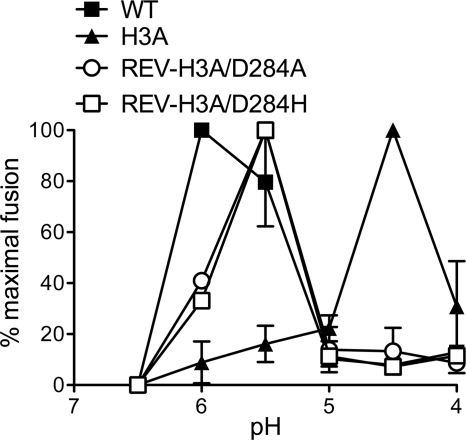
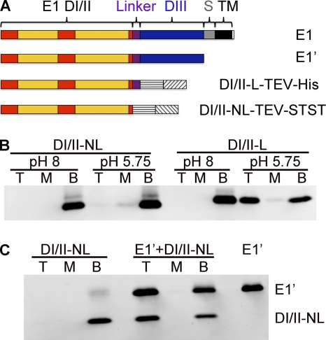
The alphavirus Semliki Forest virus (SFV) infects cells through a low-pH-dependent membrane fusion reaction mediated by the virus fusion protein E1. Acidic pH initiates a series of E1 conformational changes that culminate in membrane fusion and include dissociation of the E1/E2 heterodimer, insertion of the E1 fusion loop into the target membrane, and refolding of E1 to a stable trimeric hairpin conformation. A highly conserved histidine (H3) on the E1 protein was previously shown to promote low-pH-dependent E1 refolding. An SFV mutant with an alanine substitution at this position (H3A) has a lower pH threshold and reduced efficiency of virus fusion and E1 trimer formation than wild-type SFV. Here we addressed the mechanism by which H3 promotes E1 refolding and membrane fusion. We identified E1 mutations that rescue the H3A defect. These revertants implicated a network of interactions that connect the domain I-domain III (DI-DIII) linker region with the E1 core trimer, including H3. In support of the importance of these interactions, mutation of residues in the network resulted in more acidic pH thresholds and reduced efficiencies of membrane fusion. In vitro studies of truncated E1 proteins demonstrated that the DI-DIII linker was required for production of a stable E1 core trimer on target membranes. Together, our results suggest a critical and previously unidentified role for the DI-DIII linker region during the low-pH-dependent refolding of E1 that drives membrane fusion.


Figures

Fig. 1.
Fig. 1.
E1 trimer structure and locations of H3A second-site revertants. (A) Schematic model of the pre- and postfusion conformations of the alphavirus E1 fusion protein. The domains are colored as follows: DI in red, DII in yellow with the fusion loop as a green star, DIII in blue, the linker region between DI and DIII (DI-DIII linker) in purple, the stem in gray, and the transmembrane domain in black. The E2 protein is not shown. (B) Structure of the postfusion trimer of the E1 ectodomain (PDB accession number 1RER) (11), oriented with the fusion loops pointing to the top of the page. One chain of the trimer is colored in deep gray, another in light gray, and the third chain is colored as follows: DI in red, DII in yellow, DIII (including part of the stem) in blue, and the DI-DIII linker in purple. H3 is represented as a green stick structure. The positions of the H3A revertants are shown as space-filling structures of the parent residue, color coded as follows: P8A (lime), V10A (marine), and V140A (sky blue) in DI, and D284A/H/V (pink) and A286T (brown) in the DI-DIII linker region. A zoomed view of the region in the red box is shown in Fig. 4B. This figure was prepared with the program PyMOL (8).
Fig. 2.
Fig. 2.
pH dependence of fusion of H3A revertants. The pH dependence of membrane fusion is illustrated here for the H3A/D284A and H3A/D284H revertants, WT SFV, and the H3A mutant. Viruses were bound to BHK cells on ice for 90 min and then treated at the indicated pHs at 37°C for 1 min to trigger virus-plasma membrane fusion. Cells infected by virus fusion were quantitated by immunofluorescence assay, and the results are shown as percentages of the maximal fusion for each virus. Data are the average of two independent experiments with bars indicating the range. See Table 1 for data on other revertants.
Fig. 3.
Fig. 3.
Growth and fusion properties of D284A and D284K mutants. (A) Growth kinetics. BHK cells were electroporated with WT or mutant viral RNA and incubated at 37°C. The media containing secreted viruses were collected at the indicated times postelectroporation, and the virus titers were measured by plaque assay. (B) pH dependence of virus membrane fusion. Fusion pH dependence was measured by the fusion infection assay as described for Fig. 2. Data are the average of three determinations with bars indicating the standard deviation.
Fig. 4.
Fig. 4.
Analysis of the alphavirus E1 DI-DIII linker region. (A) Sequence alignment of the E1 DI-DIII linker region. The linker encompasses residues Pro283 to Pro294. The representative alphaviruses were selected based on the phylogenetic tree of alphaviruses (1), and the sequences were aligned using the program ClustalW. (B) Locations of the H3A reversions and the residues in the interaction network in the modified SFV E1 trimer structure. Shown is an enlarged view of the boxed region in Fig. 1B, with part of the gray chain hidden for a clear view of the interaction network. The reversion sites are shown as space-filling structures and colored in pink. The residues in the interaction network are shown as stick structures (colored and displayed as in Fig. 4C), and the backbone of the DI-DIII linker region is indicated in purple. The C0 strand on DI and the new J0 strand formed by the linker are labeled. (C) Zoom view of residues involved in the interaction network. E2 (sky blue), H3 (green), Y15 (olive), F287 (cyan), and R289 (pale green) from the interaction network are shown as stick structures. F287 and R289 are located in DI-DIII linker region shown in purple; H3 and Y15 lie in DI shown in red, and E2 is from DI of the neighboring gray chain. The potential polar interactions between E2-H3 and E2-Y15 are indicated as magenta dotted lines. Panels B and C were prepared with COOT and PyMOL (8, 9). WEEV, western equine encephalitis virus; EEEV, eastern equine encephalitis virus; VEEV, Venezuelan equine encephalitis virus.
Fig. 5.
Fig. 5.
Cell-cell fusion activities of WT and mutant SFVs. BHK cells were infected with the indicated viruses by RNA electroporation, diluted with nonelectroporated cells, and incubated at 28°C overnight to allow abundant surface expression of glycoproteins. Cell-cell fusion was triggered by treatment with buffers of the indicated pHs, and the fusion index was calculated as described in Materials and Methods. Data are the average of three determinations with bars indicating the standard deviation.
Fig. 6.
Fig. 6.
Role of the DI-DIII linker in stable membrane interaction. (A) Schematic diagrams of full-length SFV E1 and truncated E1 proteins. The E1 schematic shows the positions of the three domains (DI in red, DII in yellow, DIII in blue), with the linker (residues 283 to 294) shown in purple, the stem (S) in gray, and the transmembrane domain (TM) in black. E1′ consists of DI, DII, and DIII up to residue Pro383, followed by a double Strep tag (34). DI/II-L-TEV-His consists of DI/II-L up to Pro294, followed by a TEV cleavage site (shown as a horizontal hatched box) and a His tag (shown as an upward diagonal hatched box) (34). DI/II-NL-TEV-STST consists of DI/II-NL up to Pro283, followed by a TEV cleavage site and a double Strep tag (shown as a downward diagonal hatched box). These proteins are referred to as DI/II-NL and DI/II-L after TEV digestion. (B) DI/II-NL and DI/II-L were mixed with liposomes at final concentrations of 50 μg protein/ml and 1 mM lipid and treated at the indicated pHs for 30 min at 25°C. After neutralization, the samples were separated on sucrose flotation gradients, fractionated into the top (T), middle (M), and bottom (B) portions of the gradient, and analyzed by SDS-PAGE and Western blotting. The faint band above the major protein band is residual protein that was not cleaved by TEV. (C) DI/II-NL and E1′ (molar ratio of 1:3) were mixed with liposomes at final concentrations of 66 μg total protein/ml and 1 mM lipid. The sample was treated at pH 5.7 and analyzed as described for panel B. Panels B and C are representative examples of two experiments.
Fig. 7.
Fig. 7.
EM analysis of E1 DI/II protein-membrane interactions. The E1 DI/II-L and E1 DI/II-NL proteins were mixed with liposomes, treated at pH 5.75 for 30 min at 25°C, and analyzed by negative-stain EM. (A) Trimers are formed by the DI/II-L protein. Trimers associate in rings of 5 or 6 on the target membrane. The inset is a ×4 magnification of the boxed region and shows a ring of 6 trimers. (B) View of liposomes in the DI/II-NL protein interactions. No trimers were detected with the DI/II-NL protein. Both panels A and B are shown at a magnification of ×50,000, with the bar representing 100 nm.

Similar articles

Cited by

References

    1. Aguilar P. V., et al. 2004. Endemic Venezuelan equine encephalitis in northern Peru. Emerg. Infect. Dis. 10:880–888 - PMC - PubMed
    1. Blommel P. G., Fox B. G. 2007. A combined approach to improving large-scale production of tobacco etch virus protease. Protein Expr. Purif. 55:53–68 - PMC - PubMed
    1. Bressanelli S., et al. 2004. Structure of a flavivirus envelope glycoprotein in its low-pH-induced membrane fusion conformation. EMBO J. 23:728–738 - PMC - PubMed
    1. Centers for Disease Control and Prevention 1992. Arboviral diseases—United States, 1991. MMWR Morb. Mort. Wk. Rep. 41:545–548 - PubMed
    1. Centers for Disease Control and Prevention 1994. Arbovirus disease—United States, 1993. MMWR Morb. Mort. Wk. Rep. 43:385–387 - PubMed

Publication types

MeSH terms

LinkOut - more resources