

Oncolytic herpes simplex virus 1 encoding 15-prostaglandin dehydrogenase mitigates immune suppression and reduces ectopic primary and metastatic breast cancer in mice
- PMID: 21543507
- PMCID: PMC3126558
- DOI: 10.1128/JVI.00098-11
Oncolytic herpes simplex virus 1 encoding 15-prostaglandin dehydrogenase mitigates immune suppression and reduces ectopic primary and metastatic breast cancer in mice
Abstract
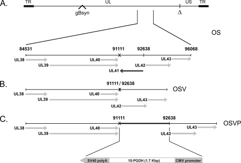
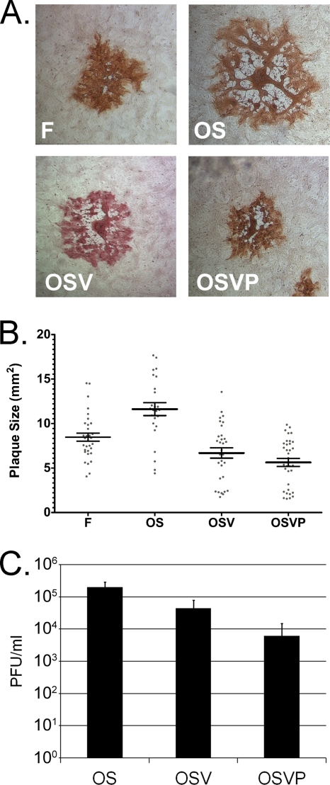
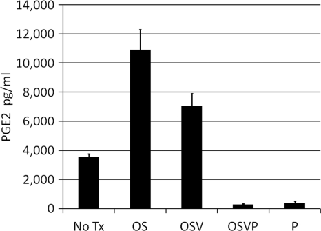
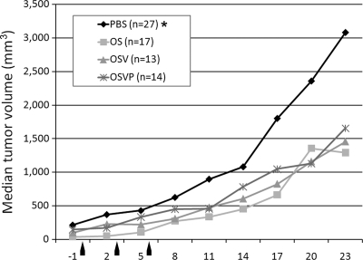
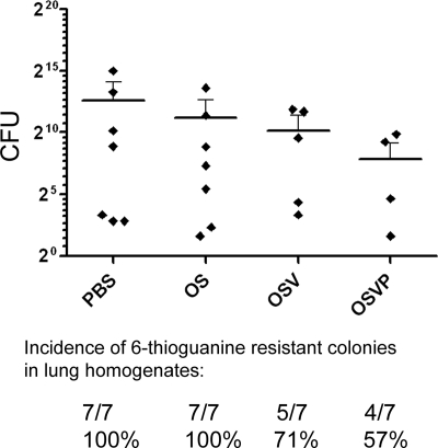
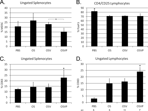
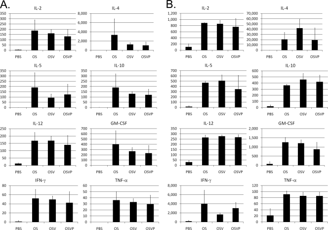
Oncolytic herpes simplex virus 1 (HSV-1) viruses armed with immunomodulatory transgenes have shown potential for enhanced antitumor therapy by overcoming tumor-based immune suppression and promoting antitumor effector cell development. Previously, we reported that the new oncolytic HSV-1 virus, OncSyn (OS), engineered to fuse tumor cells, prevented tumor growth and metastasis to distal organs in the 4T1/BALB/c immunocompetent breast cancer mouse model, suggesting the elicitation of antitumor immune responses (Israyelyan et al., Hum. Gen. Ther. 18:5, 2007, and Israyelyan et al., Virol. J. 5:68, 2008). The OSV virus was constructed by deleting the OS viral host shutoff gene (vhs; UL41) to further attenuate the virus and permit dendritic cell activation and antigen presentation. Subsequently, the OSVP virus was constructed by inserting into the OSV viral genome a murine 15-prostaglandin dehydrogenase (15-PGDH) expression cassette, designed to constitutively express 15-PGDH upon infection. 15-PGDH is a tumor suppressor protein and the primary enzyme responsible for the degradation of prostaglandin E2 (PGE2), which is known to promote tumor development. OSVP, OSV, and OS treatment of 4T1 tumors in BALB/c mice effectively reduced primary tumor growth and inhibited metastatic development of secondary tumors. OSVP was able to significantly inhibit the development and accumulation of 4T1 metastatic tumor cells in the lungs of treated mice. Ex vivo analysis of immune cells following treatment showed increased inflammatory cytokine production and the presence of mature dendritic cells for the OSVP, OSV, and OS viruses. A statistically significant decrease in splenic myeloid-derived suppressor cells (MDSC) was observed only for OSVP-treated mice. These results show that intratumoral oncolytic herpes is highly immunogenic and suggest that 15-PGDH expression by OSVP enhanced the antitumor immune response initiated by viral infection of primary tumor cells, leading to reduced development of pulmonary metastases. The availability of the OSVP genome as a bacterial artificial chromosome allows for the rapid insertion of additional immunomodulatory genes that could further assist in the induction of potent antitumor immune responses against primary and metastatic tumors.
Figures
Similar articles
-
Herpes simplex virus type-1(HSV-1) oncolytic and highly fusogenic mutants carrying the NV1020 genomic deletion effectively inhibit primary and metastatic tumors in mice.Virol J. 2008 Jun 2;5:68. doi: 10.1186/1743-422X-5-68. Virol J. 2008. PMID: 18518998 Free PMC article.
-
The Novel Oncolytic Herpes Simplex Virus Type-1 (HSV-1) Vaccine Strain VC2 Constitutively Expressing GM-CSF Causes Increased Intratumoral T Cell Infiltration and Inhibition of Tumor Metastasis in the 4T1/Balb/c Mouse Model of Stage Four Breast Cancer.J Med Virol. 2025 Feb;97(2):e70220. doi: 10.1002/jmv.70220. J Med Virol. 2025. PMID: 39930884
-
Development of a new fusion-enhanced oncolytic immunotherapy platform based on herpes simplex virus type 1.J Immunother Cancer. 2019 Aug 10;7(1):214. doi: 10.1186/s40425-019-0682-1. J Immunother Cancer. 2019. PMID: 31399043 Free PMC article.
-
Oncolytic virus therapy using genetically engineered herpes simplex viruses.Front Biosci. 2008 Jan 1;13:2060-4. doi: 10.2741/2823. Front Biosci. 2008. PMID: 17981691 Review.
-
Oncolytic herpes simplex virus type 1 and host immune responses.Curr Cancer Drug Targets. 2007 Mar;7(2):149-55. doi: 10.2174/156800907780058907. Curr Cancer Drug Targets. 2007. PMID: 17346106 Review.
Cited by
-
Oncolytic viruses: A novel treatment strategy for breast cancer.Genes Dis. 2021 Dec 16;10(2):430-446. doi: 10.1016/j.gendis.2021.11.011. eCollection 2023 Mar. Genes Dis. 2021. PMID: 37223527 Free PMC article. Review.
-
Celecoxib enhances the efficacy of 15-hydroxyprostaglandin dehydrogenase gene therapy in treating murine breast cancer.J Cancer Res Clin Oncol. 2013 May;139(5):797-807. doi: 10.1007/s00432-013-1381-9. Epub 2013 Feb 6. J Cancer Res Clin Oncol. 2013. PMID: 23385883 Free PMC article.
-
Prospects for combined use of oncolytic viruses and CAR T-cells.J Immunother Cancer. 2017 Nov 21;5(1):90. doi: 10.1186/s40425-017-0294-6. J Immunother Cancer. 2017. PMID: 29157300 Free PMC article. Review.
-
Myeloid-derived suppressor cells in breast cancer.Breast Cancer Res Treat. 2013 Jul;140(1):13-21. doi: 10.1007/s10549-013-2618-7. Epub 2013 Jul 5. Breast Cancer Res Treat. 2013. PMID: 23828498 Free PMC article. Review.
-
Myeloid-derived suppressor cells in murine retrovirus-induced AIDS inhibit T- and B-cell responses in vitro that are used to define the immunodeficiency.J Virol. 2013 Feb;87(4):2058-71. doi: 10.1128/JVI.01547-12. Epub 2012 Dec 5. J Virol. 2013. PMID: 23221564 Free PMC article.
References
-
- Aslakson C. J., Miller F. R. 1992. Selective events in the metastatic process defined by analysis of the sequential dissemination of subpopulations of a mouse mammary tumor. Cancer Res. 52:1399–1405 - PubMed
-
- Becker Y., Tavor E., Asher Y., Berkowitz C., Moyal M. 1993. Effect of herpes simplex virus type-1 UL41 gene on the stability of mRNA from the cellular genes: beta-actin, fibronectin, glucose transporter-1, and docking protein, and on virus intraperitoneal pathogenicity to newborn mice. Virus Genes 7:133–143 - PubMed
-
- Bennett J. J., et al. 2002. Comparison of safety, delivery, and efficacy of two oncolytic herpes viruses (G207 and NV1020) for peritoneal cancer. Cancer Gene Ther. 9:935–945 - PubMed
-
- Chou J., Kern E. R., Whitley R. J., Roizman B. 1990. Mapping of herpes simplex virus-1 neurovirulence to gamma 134.5, a gene nonessential for growth in culture. Science 250:1262–1266 - PubMed
Publication types
MeSH terms
Substances
Grants and funding
LinkOut - more resources
Full Text Sources
Other Literature Sources
Miscellaneous
