Comparative Study
. 2011 Aug;80(2):267-80.
doi: 10.1124/mol.110.070243. Epub 2011 May 4.

Structural and functional analysis of two new positive allosteric modulators of GluA2 desensitization and deactivation

Affiliations
Comparative Study

Structural and functional analysis of two new positive allosteric modulators of GluA2 desensitization and deactivation

David E Timm et al. Mol Pharmacol. 2011 Aug.

Abstract

At the dimer interface of the extracellular ligand-binding domain of α-amino-3-hydroxy-5-methylisoxazole-4-propionic acid (AMPA) receptors a hydrophilic pocket is formed that is known to interact with two classes of positive allosteric modulators, represented by cyclothiazide and the ampakine 2H,3H,6aH-pyrrolidino(2,1-3',2')1,3-oxazino(6',5'-5,4)benzo(e)1,4-dioxan-10-one (CX614). Here, we present structural and functional data on two new positive allosteric modulators of AMPA receptors, phenyl-1,4-bis-alkylsulfonamide (CMPDA) and phenyl-1,4-bis-carboxythiophene (CMPDB). Crystallographic data show that these compounds bind within the modulator-binding pocket and that substituents of each compound overlap with distinct moieties of cyclothiazide and CX614. The goals of the present study were to determine 1) the degree of modulation by CMPDA and CMPDB of AMPA receptor deactivation and desensitization; 2) whether these compounds are splice isoform-selective; and 3) whether predictions of mechanism of action could be inferred by comparing molecular interactions between the ligand-binding domain and each compound with those of cyclothiazide and CX614. CMPDB was found to be more isoform-selective than would be predicted from initial binding assays. It is noteworthy that these new compounds are both more potent and more effective and may be more clinically relevant than the AMPA receptor modulators described previously.


Figures

Fig. 1.
Fig. 1.
Chemical structures (top) and omit electron density maps (bottom) of two positive allosteric modulators of AMPA receptors, a, bis-alkylsulfonamide 506091 (CMPDA); b, bis-carboxythiophene 2152080 (CMPDB). Omit density map for CMPDA (a) was calculated using |Fo| − |Fc| coefficients, and view is shown perpendicular to the 2-fold axis. A simulated omit density map for CMPDB is shown (b).
Fig. 2.
Fig. 2.
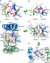
CMPDA and CMPDB cocrystals with GluA2o HS1S2H and l-glutamate. a, one molecule of CMPDA (magenta CPK) binds within the dimer interface at the clamshell hinges. View looking down the 2-fold axis. Glutamate (GLU) is shown as red CPK. HS1S2H is shown. b, view of CMPDA perpendicular to the 2-fold axis, HS1S2H is shown in surface representation. c, One molecule of CMPDB (pink CPK) binds within the dimer interface at the clamshell hinges. View looking down the 2-fold axis. Glutamate is shown as red CPK. HS1S2H is shown. d, view of CMPDB perpendicular to the 2-fold axis, HS1S2H is shown in surface representation. D1 and D2, domains 1 and 2, respectively. Coordinates have been submitted to the Protein Data Bank as 3RNN and 3RN8, respectively.
Fig. 3.
Fig. 3.
Although both modulators bind at the dimer interface and hinges, they make different contacts with the receptor. a, top view of CMPDA (magenta stick) looking down the 2-fold axis of symmetry showing the relationship of CMPDA to the hinge regions of HS1S2H. b, side view of CMPDA rotated 90° around the x-axis from view a. c, top view of CMPDB (pink stick) showing the relationship of CMPDB to the flip/flop and hinge regions of S1SJ2. d, side view of CMPDB, rotated 90° around the x-axis from view c, omitting the flip/flop regions of protomers A and B for clarity. e, side view of CMPDB rotated 90° around the y-axis from view d, omitting the hinges of protomer B for clarity. Residues within 3.2 Å of CMPDA or CMPDB are shown in ball-and-stick representation with CPK colors and carbons colored according to protomer (green, A; blue, B). Yellow residues indicate sites of point mutations. Water molecules are shown as cyan spheres. Calculated hydrogen bonds are shown as black dashed lines. Yellow dashed lines represent the Asn754–Ser729 hydrogen bond.
Fig. 4.
Fig. 4.
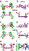
CMPDA (magenta stick, a and b) and CMPDB (pink stick, c and d) share common groups with both classes of potentiators, CTZ (green stick, left) and ampakine, CX614 (slate blue stick, right). Aside from carbons, colors are shown in CPK. Top (top) and side (bottom) views of each overlay are provided. e, alignment of CMPDA and CMPDB. f, top view of same alignment in E rotated 90° around the x-axis illustrating the position of both compounds at the hinges and flip/flop regions. Protomer A (green) and protomer B (blue) are shown in ribbon format. The coordinates for CMPDA and CMPDB in complex with the HS1S2H LBC have been submitted. The coordinates used for CTZ and CX614 were 1LBC and 2AL4, respectively.
Fig. 5.
Fig. 5.
Pharmacophores of CTZ (a), CX614 (b), CMPDA (c), and CMPDB (d) show different profiles of modulator binding. Water molecules and amino acid residues within 3.2 Å of each compound are defined by red semicircles (amino acids) or circles (water), between 3.3 and 4.9 Å (green), and greater than 5.0 Å from modulator (blue). Calculated hydrogen bonds are illustrated by black dashed arrows.
Fig. 6.
Fig. 6.
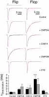
CMPDA and CMPDB modulate deactivation of GluA2 flip and flop receptors. Representative traces for homomeric WT GluA2i (left) and GluA2o (right) receptors heterologously expressed in HEK293 cells exposed to 1 ms of glutamate alone or glutamate plus each of four modulators (to measure channel deactivation). The inverted trace (outward current) above the flip control trace is a representative open-tip junction potential, which reflects the rapidity of solution exchange. Fits to the sum of two exponentials are shown in red (see Table 2). Bar plot shows mean ± S.E.M. time constant of deactivation for flip (black bars) or flop (gray bar) isoforms of rat GluA2 receptors as described under Materials and Methods.
Fig. 7.
Fig. 7.
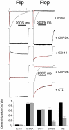
CMPDA and CMPDB modulate desensitization of GluA2 flip and flop receptors. Representative traces for homomeric WT GluA2i (left) and GluA2o (right) receptors heterologously expressed in HEK293 cells exposed to 500 ms of glutamate alone or glutamate plus each of four modulators (desensitization protocol). The inverted trace above the flip control trace is a representative open-tip junction potential indicating when glutamate was applied. Fits to the sum of two exponentials are shown in red and represent the onset of desensitization kinetics (see Table 2). Note that the calibration bar represents 200 ms for all. In addition, an expanded trace for three is shown for which the calibration bar represents 5 ms. Bar plot shows mean ± S.E.M. of the steady-state divided by peak current amplitudes (ss/pk) for flip (black bars) or flop (gray bars) GluA2 receptors.
Fig. 8.
Fig. 8.
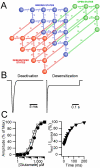
Simulations of AMPA receptor gating. a, state diagram describing a model for AMPA receptor gating in which the CC and CO are explicitly modeled. Blue circles represent closed states, green circles represent open states, and red circles represent desensitized states. Sequential glutamate binding is represented by transitions from left to right (kf and k−f) and can occur in closed open and desensitized states. Conformational transitions in the clamshell are represented by vertical transitions. Two clamshell closures are required before the transition to the open state (β and α transitions). A complete description of the states and rates of transitions is reported in the Supplemental Data (Table S1). B, simulated responses to 1- and 500-ms pulses of 10 mM glutamate. A single exponential fit of the decays results in a τDeact = 0.7 ms and τDes = 6.2 ms, similar to GluA2i experimental data. C, simulated dose response for peak (●) and steady-state (□) currents in response to 1 to 10,000 μM pulses of glutamate (left); simulated recovery from desensitization in a paired pulse paradigm (right). Fits to the simulated data yield EC50peak = 292 μM, EC50ss = 139 μM, and a τrecovery = 59 ms. These fits are close approximations to experimental data presented in this article and to data that have been reported previously (Zhang et al., 2008).
Fig. 9.
Fig. 9.
Changes in rate constants in the model predict changes in the kinetics of deactivation and desensitization observed by modulators. Simulations of GluA2 currents under voltage-clamp in response to 10 mM glutamate for either a 1-ms pulse to observe deactivation kinetics (top) or a 500-ms pulse to observe desensitization kinetics (bottom). Simulated GluA2 currents under control conditions (no modulator) are represented by gray traces. Potential effects of modulator (black traces) are segregated by column and have been simulated by slowing either the CO, α, or δ transitions in isolation or in combination. The middle row shows how changes in the rate constants influence the stability of various receptor states, as measured by estimated changes in Gibb's free energy (ΔG). The troughs from left to right represent the following: the unbound receptor, the agonist bound receptor in the open clamshell state, the agonist bound receptor in the closed clamshell state, and the open channel. The lower middle row repeats the trough found above it and shows the transition to the agonist bound desensitized state (states not shown are identical with those shown above). Results indicate that CTZ can be simulated by slowing δ, CMPDB can be simulated by slowing α, and CMPDA can be simulated by slowing α and δ or CO and δ.

References

    1. Ahmed AH, Wang Q, Sondermann H, Oswald RE. (2009) Structure of the S1S2 glutamate binding domain of GLuR3. Proteins 75:628–637 - PMC - PubMed
    1. Arai A, Kessler M, Ambros-Ingerson J, Quan A, Yigiter E, Rogers G, Lynch G. (1996a) Effects of a centrally active benzoylpyrrolidine drug on AMPA receptor kinetics. Neuroscience 75:573–585 - PubMed
    1. Arai A, Kessler M, Rogers G, Lynch G. (1996b) Effects of a memory-enhancing drug on DL-alpha-amino-3-hydroxy-5-methyl-4-isoxazolepropionic acid receptor currents and synaptic transmission in hippocampus. J Pharmacol Exp Ther 278:627–638 - PubMed
    1. Arai AC, Kessler M, Rogers G, Lynch G. (2000) Effects of the potent ampakine CX614 on hippocampal and recombinant AMPA receptors: interactions with cyclothiazide and GYKI 52466. Mol Pharmacol 58:802–813 - PubMed
    1. Arai AC, Xia YF, Rogers G, Lynch G, Kessler M. (2002) Benzamide-type AMPA receptor modulators form two subfamilies with distinct modes of action. J Pharmacol Exp Ther 303:1075–1085 - PubMed

Publication types

Substances

LinkOut - more resources