

Pulmonary pathology of pandemic influenza A/H1N1 virus (2009)-infected ferrets upon longitudinal evaluation by computed tomography
- PMID: 21543558
- PMCID: PMC3167882
- DOI: 10.1099/vir.0.032805-0
Pulmonary pathology of pandemic influenza A/H1N1 virus (2009)-infected ferrets upon longitudinal evaluation by computed tomography
Abstract
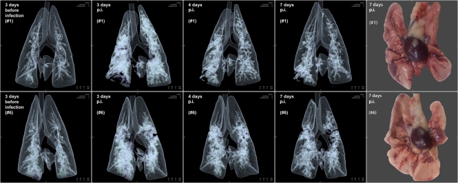
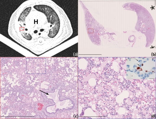
We investigated the development of pulmonary lesions in ferrets by means of computed tomography (CT) following infection with the 2009 pandemic A/H1N1 influenza virus and compared the scans with gross pathology, histopathology and immunohistochemistry. Ground-glass opacities observed by CT scanning in all infected lungs corresponded to areas of alveolar oedema at necropsy. These areas were most pronounced on day 3 and gradually decreased from days 4 to 7 post-infection. This pilot study shows that the non-invasive imaging procedure allows quantification and characterization of influenza-induced pulmonary lesions in living animals under biosafety level 3 conditions and can thus be used in pre-clinical pharmaceutical efficacy studies.
Figures
Similar articles
-
Molecular imaging reveals a progressive pulmonary inflammation in lower airways in ferrets infected with 2009 H1N1 pandemic influenza virus.PLoS One. 2012;7(7):e40094. doi: 10.1371/journal.pone.0040094. Epub 2012 Jul 20. PLoS One. 2012. PMID: 22911695 Free PMC article.
-
Clinical response to pandemic H1N1 influenza virus from a fatal and mild case in ferrets.Virol J. 2015 Mar 26;12:48. doi: 10.1186/s12985-015-0272-x. Virol J. 2015. PMID: 25888921 Free PMC article.
-
Key molecular factors in hemagglutinin and PB2 contribute to efficient transmission of the 2009 H1N1 pandemic influenza virus.J Virol. 2012 Sep;86(18):9666-74. doi: 10.1128/JVI.00958-12. Epub 2012 Jun 27. J Virol. 2012. PMID: 22740390 Free PMC article.
-
Comparison of the pathology caused by H1N1, H5N1, and H3N2 influenza viruses.Arch Med Res. 2009 Nov;40(8):655-61. doi: 10.1016/j.arcmed.2009.10.001. Epub 2010 Jan 6. Arch Med Res. 2009. PMID: 20304252 Review.
-
Swine-origin influenza A (H1N1) viral infection: thoracic findings on CT.AJR Am J Roentgenol. 2011 Jun;196(6):W723-8. doi: 10.2214/AJR.10.5109. AJR Am J Roentgenol. 2011. PMID: 21606260 Review.
Cited by
-
The use of nonhuman primates in research on seasonal, pandemic and avian influenza, 1893-2014.Antiviral Res. 2015 May;117:75-98. doi: 10.1016/j.antiviral.2015.02.011. Epub 2015 Mar 5. Antiviral Res. 2015. PMID: 25746173 Free PMC article. Review.
-
Molecular imaging reveals a progressive pulmonary inflammation in lower airways in ferrets infected with 2009 H1N1 pandemic influenza virus.PLoS One. 2012;7(7):e40094. doi: 10.1371/journal.pone.0040094. Epub 2012 Jul 20. PLoS One. 2012. PMID: 22911695 Free PMC article.
-
Quantitative measurement of influenza virus replication using consecutive bronchoalveolar lavage in the lower respiratory tract of a ferret model.J Vet Sci. 2014;15(3):439-42. doi: 10.4142/jvs.2014.15.3.439. Epub 2014 Apr 2. J Vet Sci. 2014. PMID: 24690606 Free PMC article.
References
-
- Baras B., de Waal L., Stittelaar K. J., Jacob V., Giannini S., Veldhuis Kroeze E. J., van den Brand J. M. A., van Amerongen G., Simon J. H., et al. (2011). Pandemic H1N1 vaccine requires the use of an adjuvant to protect against challenge in naïve ferrets. Vaccine 29, 2120–2126 10.1016/j.vaccine.2010.12.125 - DOI - PubMed
-
- CDC (2009). Update: infections with a swine-origin influenza A (H1N1) virus–United States and other countries, April 28, 2009. MMWR Morb Mortal Wkly Rep 58, 431–433 - PubMed
-
- Del Giudice G., Stittelaar K. J., van Amerongen G., Simon J., Osterhaus A. D., Stöhr K., Rappuoli R. (2009). Seasonal influenza vaccine provides priming for A/H1N1 immunization. Sci Transl Med 1, re1. - PubMed
-
- Friesen R. H., Koudstaal W., Koldijk M. H., Weverling G. J., Brakenhoff J. P., Lenting P. J., Stittelaar K. J., Osterhaus A. D., Kompier R., Goudsmit J. (2010). New class of monoclonal antibodies against severe influenza: prophylactic and therapeutic efficacy in ferrets. PLoS ONE 5, e9106 10.1371/journal.pone.0009106 - DOI - PMC - PubMed
Publication types
MeSH terms
LinkOut - more resources
Full Text Sources
Medical