. 2011 May 4;31(18):6850-7.
doi: 10.1523/JNEUROSCI.6467-10.2011.

BACE1 activity is modulated by cell-associated sphingosine-1-phosphate

Affiliations

BACE1 activity is modulated by cell-associated sphingosine-1-phosphate

Nobumasa Takasugi et al. J Neurosci. .

Abstract

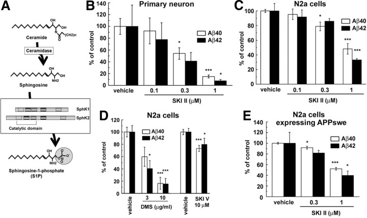
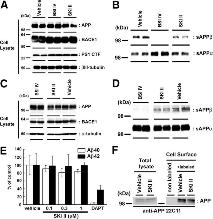
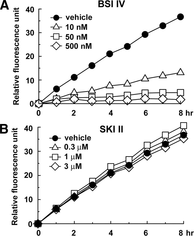
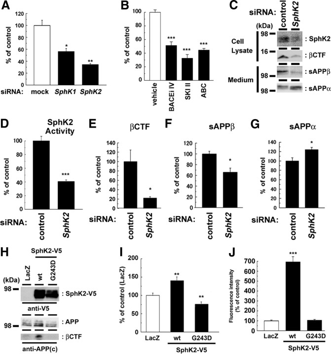
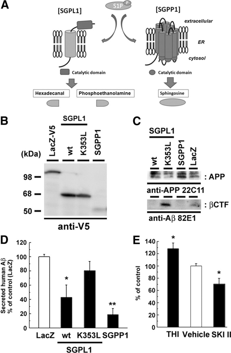
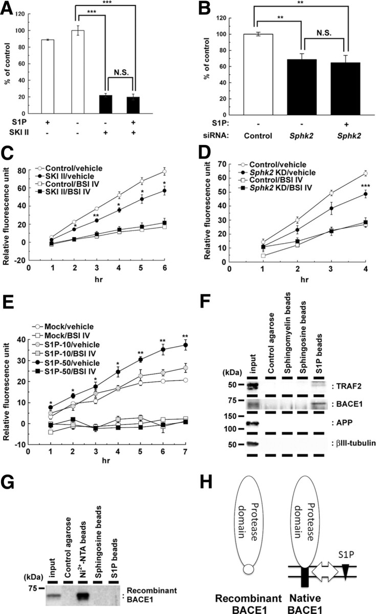
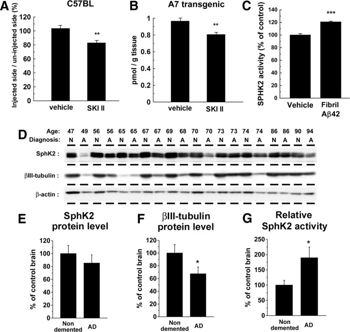
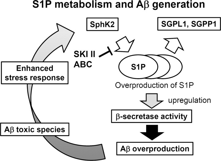
Sphingosine kinase (SphK) 1 and 2 phosphorylate sphingosine to generate sphingosine-1-phosphate (S1P), a pluripotent lipophilic mediator implicated in a variety of cellular events. Here we show that the activity of β-site APP cleaving enzyme-1 (BACE1), the rate-limiting enzyme for amyloid-β peptide (Aβ) production, is modulated by S1P in mouse neurons. Treatment by SphK inhibitor, RNA interference knockdown of SphK, or overexpression of S1P degrading enzymes decreased BACE1 activity, which reduced Aβ production. S1P specifically bound to full-length BACE1 and increased its proteolytic activity, suggesting that cellular S1P directly modulates BACE1 activity. Notably, the relative activity of SphK2 was upregulated in the brains of patients with Alzheimer's disease. The unique modulatory effect of cellular S1P on BACE1 activity is a novel potential therapeutic target for Alzheimer's disease.


Figures

Figure 1.
Figure 1.
Sphingosine kinase inhibitors decreased the Aβ secretion from neuronal cells. A, Schematic depiction of synthetic pathway for S1P. Five conserved regions of SphK are indicated by rectangles. Location of catalytic region is also indicated in this diagram. B–E, Effects of SKI on secretion of Aβ40 and Aβ42 from neuronal cells. The levels of secreted Aβ in conditioned media were quantified by ELISAs. Mean ± SEM percentages of the relative ratio of secreted Aβ to levels in untreated control are indicated. *p < 0.05, ***p < 0.001 by Student's t test. B, Levels of secreted Aβ from mouse primary cortical neurons (7 d in vitro) after treatment with SKI II for 24 h (n = 4). C, Levels of secreted Aβ from N2a cells after treatment with SKI II for 24 h (n = 4). D, Levels of secreted Aβs from N2a cells after treatment with DMS or SKI V for 24 h (n = 4–7). E, Levels of secreted Aβ measured by human Aβ-specific ELISA from N2a cells stably expressing Swedish mutant of APP after treatment with SKI II for 24 h (n = 4).
Figure 2.
Figure 2.
SKI II decreased the β-secretase cleavage products. A–D, Immunoblot analyses of the protein levels of APP derivatives and BACE1 in neuronal cells. Immunoblot analysis of cell lysates (A) and cultured media (B) of mouse primary cortical neurons (7 d in vitro) treated with BACE inhibitor IV (BSI IV; 1 μm) or SKI II (1 μm) for 24 h in duplicate (n = 3; representative results are shown). Immunoblot analysis of cell lysates (C) and cultured media (D) of naive N2a cells treated with BACE inhibitor IV (BSI IV; 1 μm) or SKI II (3 μm) for 24 h. E, Levels of secreted human Aβ from N2a cells overexpressing SC100 after treatment with SKI II for 24 h. Secreted human Aβ40 and Aβ42 were detected by human Aβ-specific ELISA (n = 4; mean ± SEM). F, Effect of SKI II (1 μm) on the cell-surface levels of APP in naive N2a cells. After treatment with vehicle or SKI II for 24 h, N2a cells were biotinylated by sulfo-NHS-biotin and pulled down by streptavidin beads.
Figure 3.
Figure 3.
Effect of SKI II on the catalytic activity of BACE1. In vitro BACE1 activity assay using a fluorogenic BACE1-specific substrate. BACE inhibitor IV (BSI IV; A) or SKI II (B) was coincubated with recombinant soluble BACE1 protein at indicated duration and concentrations. Relative fluorescence units were shown (n = 3).
Figure 4.
Figure 4.
SphK2 activity modulated the β-secretase cleavage products. A, N2a cells were transiently transfected with siRNAs against endogenous SphKs. After 48 h transfection, media were replaced and further incubated for 24 h. Levels of secreted Aβ were quantified by ELISA (n = 3; mean ± SEM; *p < 0.05, **p < 0.01). B, Levels of secreted Aβ from N2a cells treated with BACE inhibitor IV (BACEi IV), SKI II, or SphK2-selective inhibitor ABC294640 (ABC) for 24 h (n = 3; mean ± SEM; ***p < 0.001). C–G, Effect of transient SphK2 knockdown on APP derivatives in N2a cells. Representative immunoblot analysis was shown in C. In vitro SphK2 activity (D) as well as βCTF (E) in cell lysates and the amount of sAPPβ in conditioned media (F) were significantly decreased by knockdown of SphK2. In contrast, the level of sAPPα in conditioned media (G) was significantly increased (quantitated by densitometric analysis; n = 4; mean ± SEM; *p < 0.05). H–J, Effect of SphK2 on N2a cells coexpressing Swedish mutant of APP. wt, Wild type. Representative immunoblot analysis was shown in H. Overexpression of SphK2, but not inactive mutant (G243D), increased the levels of Aβ production (I) as well as SphK2 activity in vitro (J) (n = 4; mean ± SEM; *p < 0.05, **p < 0.01).
Figure 5.
Figure 5.
Effect of S1P degrading enzymes on Aβ production. A, Schematic view of S1P degradation pathway. Note that SGPL1 and SGPP1 generate different degradation products of S1P. ER, Endoplasmic reticulum. B–D, Effects of the overexpression of V5-tagged S1P degrading enzymes on APP metabolism in N2a cells. N2a cells were cotransfected with S1P degrading enzymes and Swedish mutant of APP. After 24 h transfection, media were replaced and further incubated for 24 h. Immunoblot analysis of S1P degrading enzymes (B) and APP derivatives (C) are shown. Human APP-derived βCTF was specifically detected by an anti-human Aβ N-terminus antibody (82E1). wt, Wild type. D, The levels of secreted human Aβ was detected by human Aβ-specific ELISA (n = 4; mean ± SEM; *p < 0.05, **p < 0.01). Note that overexpression of SGPL1 or SGPP1, but not SGPL1 carrying catalytically inactive mutation (K353L), decreased the generation of βCTF and the Aβ secretion from N2a cells. E, The levels of secreted Aβ from mouse primary cortical neurons (7 d in vitro) treated with SGPL1 inhibitor THI (50 μg/ml) or SKI II (1 μm) for 24 h (n = 4; mean ± SEM; *p < 0.05).
Figure 6.
Figure 6.
SKI II treatment decreased the β-secretase activity in cellular membrane. A, B, Effect of extracellularly added S1P (10 μm) on levels of secreted Aβ from mouse primary cortical neurons (7 d in vitro) after treatment with SKI II (1 μm) for 24 h (A) (n = 4; mean ± SEM) or from N2a cells after 48 h SphK2 knockdown (B) (n = 3; mean ± SEM; **p < 0.01, ***p < 0.001; N.S., no significant difference). Note that S1P failed to rescue the decrease in Aβ production either by SKI II or SphK2 knockdown. C, β-Secretase activity in the membrane fractions of N2a cells treated with vehicle or SKI II (1 μm) for 24 h. BACE inhibitor IV (BSI IV; 1 μm) was added to the in vitro assay (n = 3; mean ± SEM; *p < 0.05, **p < 0.01 vs control/vehicle). D, β-Secretase activity in the membrane fractions of SphK2 knockdown N2a cells. BACE inhibitor IV (BSI IV; 1 μm) was added to the in vitro assay (n = 3; mean ± SEM; ***p < 0.001 vs control/vehicle). E, Effect of S1P on β-secretase activity in the membrane fractions of mouse brain. S1P (10 or 50 μm) and BACE inhibitor IV (BSI IV; 1 μm) were added to the in vitro assay (n = 3; mean ± SEM; *p < 0.05, **p < 0.01 vs mock/vehicle). F, G, Association of BACE1 holoprotein with immobilized S1P. N2a cell lysates (F) or recombinant BACE1 with 10×His tag that lacks the transmembrane and cytoplasmic domains (G) were incubated with control agarose (no lipid), Nickel-NTA agarose, sphingomyelin, sphingosine, or S1P-coated affinity matrices (as indicated), and bound proteins were analyzed by immunoblotting. H, Schematic model of the binding of BACE1 and S1P. S1P (black triangles) interacts with the C-terminal region of BACE1 (black squares), including the transmembrane domain, but not with the extracellular protease domain (white ovals). Location of 10×His tag is indicated by a white circle.
Figure 7.
Figure 7.
Role of SphK2 activity in AD brains. A, Effect of the direct injection of SKI II into the hippocampus of nontransgenic wild-type female mice (C57BL) at 8 weeks of age. The levels of Tris-soluble Aβ in the injected side of hippocampus were divided by those in the uninjected side. Data represent relative ratio of each group (n = 4; mean ± SEM; **p < 0.01). B, Levels of Tris-soluble Aβ in the cerebral cortices of female A7 mice at 6 months of age after a 7 d treatment with SKI II (50 mg · kg−1 · d−1, p.o.). Total brain Aβ levels were measured by human-specific sandwich ELISA (n = 5; mean ± SEM; **p < 0.01). C, Effect of Aβ fibril on SphK2 activity in N2a cells. N2a cells were treated with Aβ42 fibril (30 μm) overnight, and cell lysates were subjected to an in vitro SphK2 activity assay (n = 3; mean ± SEM; ***p < 0.001). D–F, Immunoblot analysis of Tris-soluble fractions (15 μg of protein in each lane) from cortices of AD (denoted as A) or non-demented (denoted as N) individuals. Average protein levels of SphK2 (E) and βIII-tubulin (F) in each individual were analyzed by densitometric analyses (*p < 0.05). G, Average of in vitro SphK2 enzymatic activity of Tris-soluble fractions from brains of AD and non-demented individuals. The enzymatic activities of SphK2 were normalized by the protein levels of SphK2 quantified in D.
Figure 8.
Figure 8.
Schematic representation of the role of S1P metabolism in AD.

Similar articles

Cited by

References

    1. Ahmed RR, Holler CJ, Webb RL, Li F, Beckett TL, Murphy MP. BACE1 and BACE2 enzymatic activities in Alzheimer's disease. J Neurochem. 2010;112:1045–1053. - PMC - PubMed
    1. Alvarez SE, Milstien S, Spiegel S. Autocrine and paracrine roles of sphingosine-1-phosphate. Trends Endocrinol Metab. 2007;18:300–307. - PubMed
    1. Alvarez SE, Harikumar KB, Hait NC, Allegood J, Strub GM, Kim EY, Maceyka M, Jiang H, Luo C, Kordula T, Milstien S, Spiegel S. Sphingosine-1-phosphate is a missing cofactor for the E3 ubiquitin ligase TRAF2. Nature. 2010;465:1084–1088. - PMC - PubMed
    1. Arnold SE, Lee EB, Moberg PJ, Stutzbach L, Kazi H, Han LY, Lee VM, Trojanowski JQ. Olfactory epithelium amyloid-β and paired helical filament-tau pathology in Alzheimer disease. Ann Neurol. 2010;67:462–469. - PMC - PubMed
    1. Blondeau N, Lai Y, Tyndall S, Popolo M, Topalkara K, Pru JK, Zhang L, Kim H, Liao JK, Ding K, Waeber C. Distribution of sphingosine kinase activity and mRNA in rodent brain. J Neurochem. 2007;103:509–517. - PMC - PubMed

Publication types