

Relationship between surface area of nonperfused myocardium and extravascular extraction of contrast agent following coronary microembolization
- PMID: 21543631
- PMCID: PMC3154707
- DOI: 10.1152/ajpregu.00428.2010
Relationship between surface area of nonperfused myocardium and extravascular extraction of contrast agent following coronary microembolization
Abstract
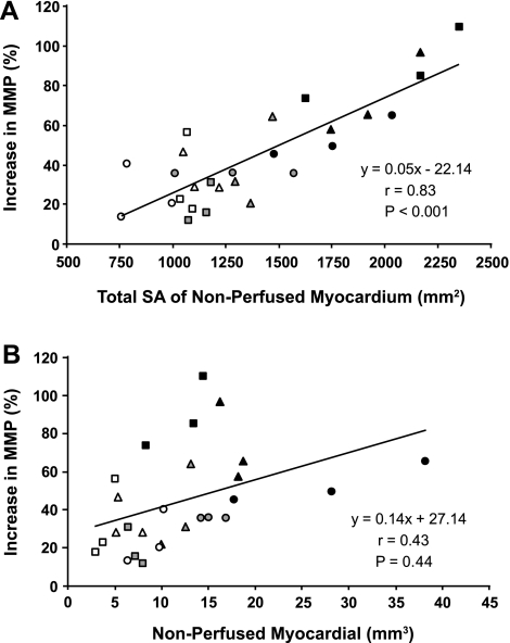
Myocardial microvascular permeability and coronary sinus concentration of muscle metabolites have been shown to increase after myocardial ischemia due to epicardial coronary artery occlusion and reperfusion. However, their association with coronary microembolization is not well defined. This study tested the hypothesis that acute coronary microembolization increases microvascular permeability in the porcine heart. The left anterior descending perfusion territories of 34 anesthetized pigs (32 ± 3 kg) were embolized with equal volumes of microspheres of one of three diameters (10, 30, or 100 μm) and at three different doses for each size. Electron beam computed tomography (EBCT) was used to assess in vivo, microvascular extraction of a nonionic contrast agent (an index of microvascular permeability) before and after microembolization with microspheres at baseline and during adenosine infusion. A high-resolution three-dimensional microcomputed tomography (micro-CT) scanner was subsequently used to obtain ex vivo, the volume and corresponding surface area of the embolized myocardial islands within the perfusion territories of the microembolized coronary artery. EBCT-derived microvascular extraction of contrast agent increased within minutes after coronary microembolization (P < 0.001 vs. baseline and vs. control values). The increase in coronary microvascular permeability was highly correlated to the micro-CT-derived total surface area of the nonperfused myocardium (r = 0.83, P < 0.001). In conclusion, myocardial extravascular accumulation of contrast agent is markedly increased after coronary microembolization and its magnitude is in proportion to the surface area of the interface between the nonperfused and perfused territories.
Figures


References
-
- Albers J, Schroeder A, de Simone R, Mockel R, Vahl CF, Hagl S. 3D evaluation of myocardial edema: experimental study on 22 pigs using magnetic resonance and tissue analysis. Thorac Cardiovasc Surg 49: 199–203, 2001 - PubMed
-
- Angelini A, Rubartelli P, Mistrorigo F, Della Barbera M, Abbadessa F, Vischi M, Thiene G, Chierchia S. Distal protection with a filter device during coronary stenting in patients with stable and unstable angina. Circulation 110: 515–521, 2004 - PubMed
-
- Beighley PE, Thomas PJ, Jorgensen SM, Ritman EL. 3D architecture of myocardial microcirculation in intact rat heart: a study with micro-CT. Adv Exp Med Biol 430: 165–175, 1997 - PubMed
-
- Belmin J, Corman B, Merval R, Tedgui A. Age-related changes in endothelial permeability and distribution volume of albumin in rat aorta. Am J Physiol Heart Circ Physiol 264: H679–H685, 1993 - PubMed
Publication types
MeSH terms
Substances
Grants and funding
LinkOut - more resources
Full Text Sources
Medical