. 2011 Aug 4;118(5):1305-15.
doi: 10.1182/blood-2011-01-331462. Epub 2011 May 4.

Toll-like receptor 7 (TLR7)-driven accumulation of a novel CD11c⁺ B-cell population is important for the development of autoimmunity

Affiliations

Toll-like receptor 7 (TLR7)-driven accumulation of a novel CD11c⁺ B-cell population is important for the development of autoimmunity

Anatoly V Rubtsov et al. Blood. .

Abstract

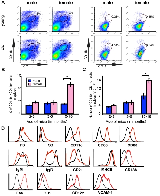
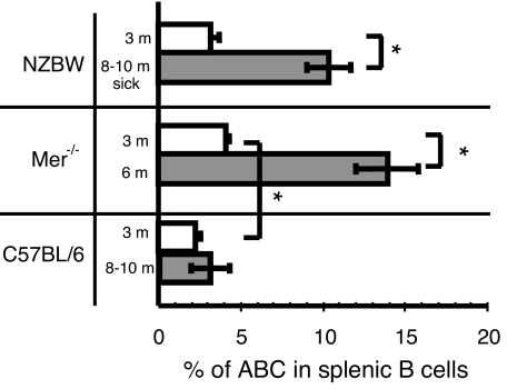
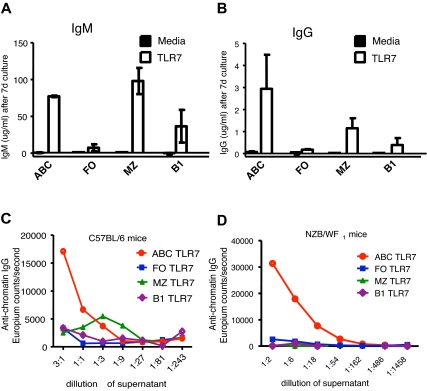
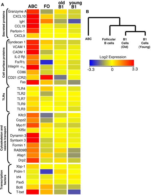
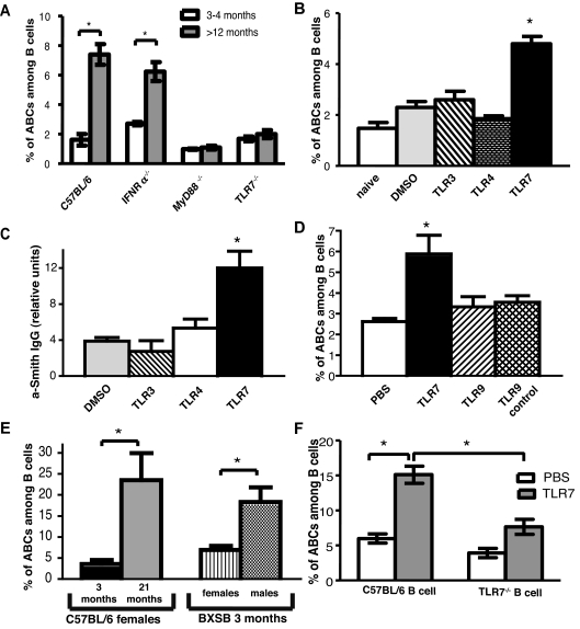
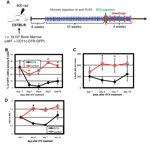
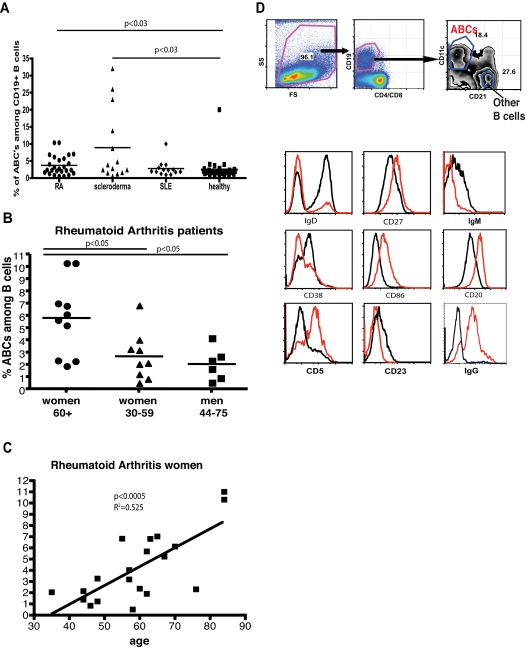
Females are more susceptible than males to many autoimmune diseases. The processes causing this phenomenon are incompletely understood. Here, we demonstrate that aged female mice acquire a previously uncharacterized population of B cells that we call age-associated B cells (ABCs) and that these cells express integrin α(X) chain (CD11c). This unexpected population also appears in young lupus-prone mice. On stimulation, CD11c(+) B cells, both from autoimmune-prone and healthy strains of mice, secrete autoantibodies, and depletion of these cells in vivo leads to reduction of autoreactive antibodies, suggesting that the cells might have a direct role in the development of autoimmunity. We have explored factors that contribute to appearance of ABCs and demonstrated that signaling through Toll-like receptor 7 is crucial for development of this B cell population. We were able to detect a similar population of B cells in the peripheral blood of some elderly women with autoimmune disease, suggesting that there may be parallels between the creation of ABC-like cells between mice and humans.


Figures

Figure 1
Figure 1
Elderly female mice contain an enlarged population of CD19+CD11b+CD11c+ B cells (ABCs). (A) Flow cytometric analysis of total spleen (left set of plots) or splenic B cells (gated as IgM+B220+CD4CD8NK1.1; right set of plots) in young (< 12 weeks old) and elderly (> 1 year old) C57BL/6 mice. Data are representative of > 10 independent analyses. (B-C) Average percent and number of CD19+CD11b+CD11c+ cells in spleen of male (blue) and female (pink) C57BL/6 mice. *P < .01 (Student 2-tailed t test). (D) Flow cytometry of FO B cells (CD19+CD11b; black) and CD19+CD11b+ B cells (red).
Figure 2
Figure 2
Increased number of ABCs in autoimmune prone mice at the time of onset of autoimmunity. The percentage of ABCs in splenic B cell populations was determined by flow cytometry in female mice of indicated strain and age. Bars represent mean (± SEM) of at least 5 mice per group. *P < .01 (Student 2-tailed t test).
Figure 3
Figure 3
ABCs produce anti-chromatin antibodies on stimulation in vitro. ABCs and FO, MZ, and B1 B cells were isolated from C57BL/6 (A-C) or NZB/WF1 (D) mice and cultured for 7 days in the presence of medium or TLR7 agonist. Total IgM (A), IgG (B), and anti-chromatin IgG (C-D) were subsequently measured in supernatant by enzyme-linked immunosorbent assay. (A-B) Bars represent mean (± SEM) of 3 independent experiments. (C-D) Data are representative of 3 independent experiments.
Figure 4
Figure 4
Transcriptome analysis of ABCs and FO, MZ, and B1 B cells. (A) Results for some of the genes that changed expression level only in ABCs, together with expression values for some control genes. Up- and down- regulated transcripts are indicated in red and blue, respectively. The magnitude of expression is depicted by the color bar. (B) A genealogical tree, based on gene expression by the different B cell populations that were analyzed, was created by GeneSpring X software based on gene expression profile of analyzed B cell populations.
Figure 5
Figure 5
TLR7 and MyD88 signaling is required for ABC accumulation. (A) Average percentage of ABCs among B cell in spleen of young (12-16 weeks old) and aged (> 12 months old) C57BL/6, IFNR−/−, and TLR7−/−, MyD88−/− female mice. (B) Average percentage of ABCs among B cells in spleen of young (8-12 weeks old) C57BL/6 female mice after 30 immunizations with vehicle or the indicated TLR agonist. (C) Anti-Smith IgG autoantibodies were measured by ELISA in serum of mice chronically injected with TLR agonists. (D) Average percentage of ABCs among B cells in spleen of young (8-12 weeks old) C57BL/6 female mice after 30 immunizations with indicated TLR agonist. (E) Average percentage of ABCs among B cells in spleen of young (12-week-old) BXSB male and female mice. (F) Bone marrow chimeras possessing wild-type or TLR7−/− B cells were treated with vehicle or TLR7 agonist for 2 months, and the percentage of ABCs in the spleen was determined by flow cytometric analysis. Bars represent mean (± SEM) of at least 10 mice per group. *P < .05 (Student 2-tailed t test).
Figure 6
Figure 6
Depletion of ABCs reduces amount of autoantibodies in serum. Bone marrow chimeras were constructed as described under “Generation of bone marrow chimeras” such that the only CD11c population that entirely expressed the DTR was that of the ABCs. The chimeras were injected 3 times a week with TLR7 agonist, and 8 to 12 weeks later, they were analyzed for their titer of anti-Smith antibodies. The mice were assigned to control and DT-treated groups by matching mice between the groups for their titers of anti-Smith antibodies and then injected as controls or with DT. (A) Schematic outline of the experiment. (B) Percentage of ABCs in blood before and after injection of DT. (C) Anti-Smith IgG autoantibodies were measured by ELISA in mouse serum at various times after injection of DT. (D) Data presented in Figure 7C are normalized to titers of anti-Smith antibodies of day 1. Bars in panels B through D represent mean (± SEM) of at least 5 mice per group. Data are representative of 2 independent analyses. *P < .05 (Student 2-tailed t test).
Figure 7
Figure 7
Presence of ABC-like cells in human peripheral blood. (A) Percentage of ABC-like cells among CD19+ cells in the peripheral blood of patients with RA, SSc, or SLE, or in HDs. Statistical analysis performed using the Mann-Whitney test. (B) Percentage of ABC-like cells among CD19+ cells in peripheral blood of women > 60 years old, women < 60 years old, or men among patients with RA. Statistical analysis performed using the Mann-Whitney test. (C) Correlation of percentage of ABCs among CD19+ B cells in blood with the age of female patients with RA. (D) Phenotypic characterization of human ABCs (red) and other B cells (black). Gating strategy is shown at the top part of the figure.

Comment in

  • Now you know your ABCs.
    Pillai S. Pillai S. Blood. 2011 Aug 4;118(5):1187-8. doi: 10.1182/blood-2011-06-355131. Blood. 2011. PMID: 21816835 No abstract available.

Similar articles

Cited by

References

    1. Beeson PB. Age and sex associations of 40 autoimmune diseases. Am J Med. 1994;96(5):457–462. - PubMed
    1. Whitacre CC. Sex differences in autoimmune disease. Nat Immunol. 2001;2(9):777–780. - PubMed
    1. Kennedy LG, Will R, Calin A. Sex ratio in the spondyloarthropathies and its relationship to phenotypic expression, mode of inheritance and age at onset. J Rheumatol. 1993;20(11):1900–1904. - PubMed
    1. Roubinian JR, Papoian R, Talal N. Androgenic hormones modulate autoantibody responses and improve survival in murine lupus. J Clin Invest. 1977;59(6):1066–1070. - PMC - PubMed
    1. Gubbels Bupp MR, Jorgensen TN, Kotzin BL. Identification of candidate genes that influence sex hormone-dependent disease phenotypes in mouse lupus. Genes Immun. 2008;9(1):47–56. - PubMed

Publication types

MeSH terms