. 2011 Sep;85(3):594-604.
doi: 10.1095/biolreprod.110.088542. Epub 2011 May 4.

Mice lacking the USP2 deubiquitinating enzyme have severe male subfertility associated with defects in fertilization and sperm motility

Affiliations

Mice lacking the USP2 deubiquitinating enzyme have severe male subfertility associated with defects in fertilization and sperm motility

Nathalie Bedard et al. Biol Reprod. 2011 Sep.

Abstract

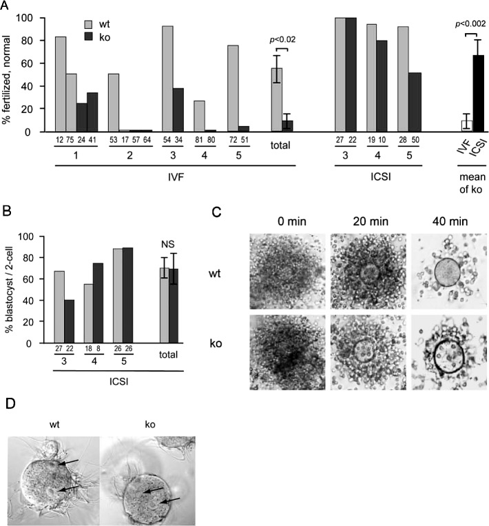
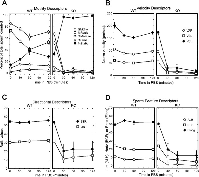
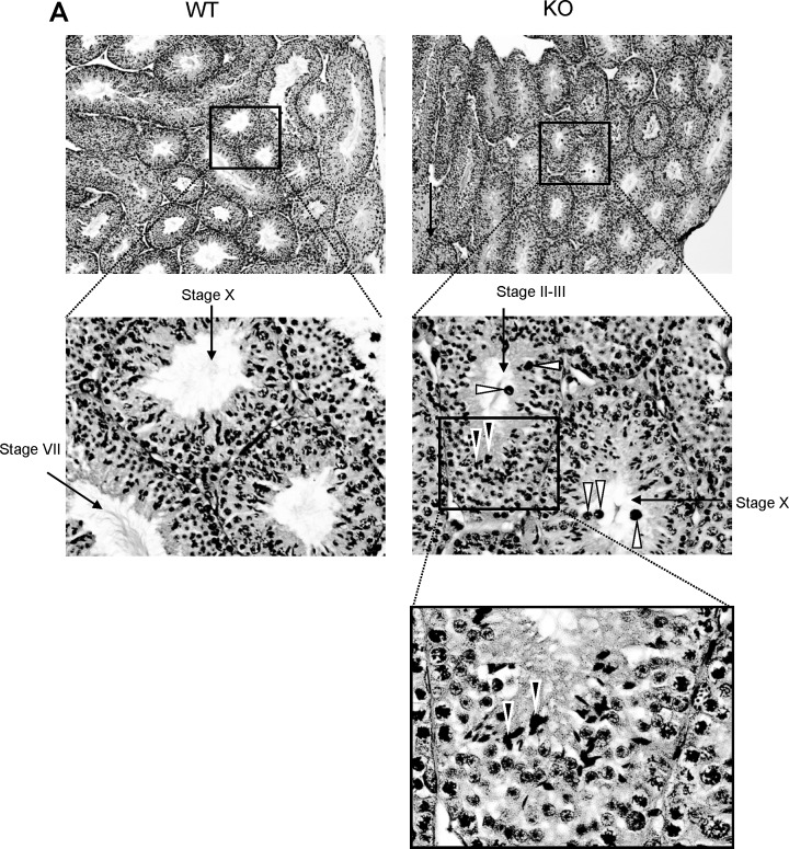
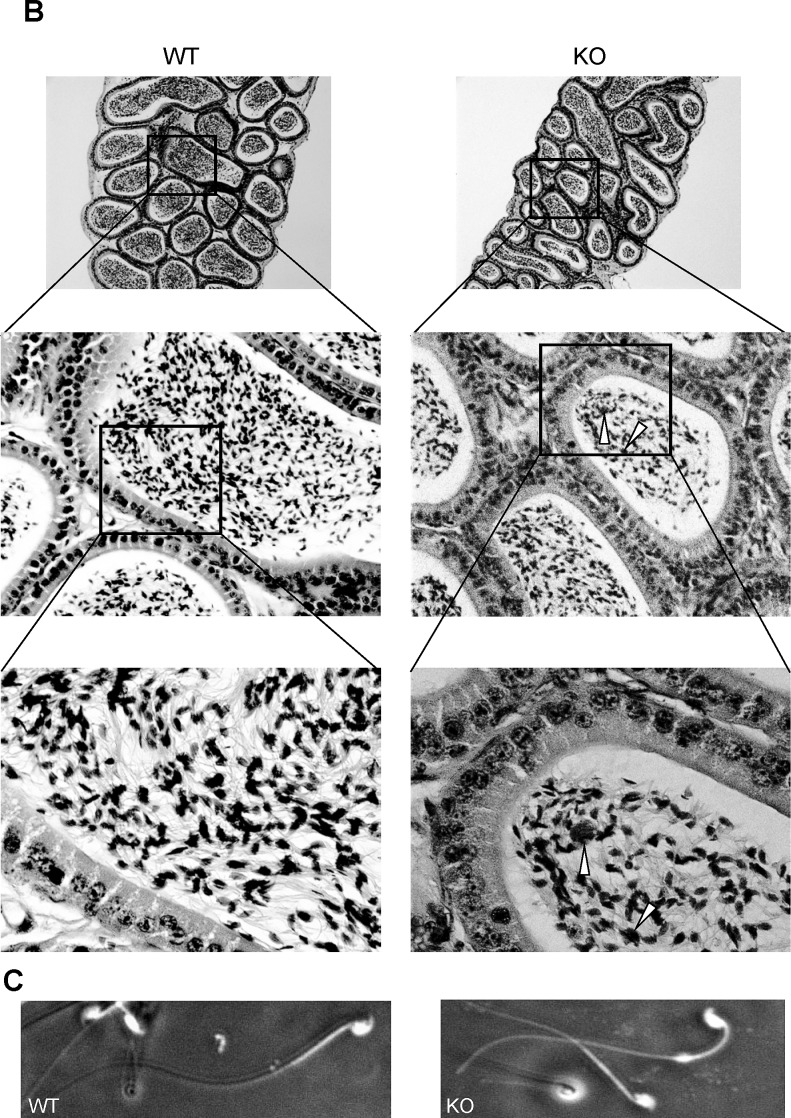
The ubiquitin-proteasome system plays an important role in spermatogenesis. However, the functions of deubiquitinating enzymes in this process remain poorly characterized. We previously showed that the deubiquitinating enzyme USP2 is induced in late elongating spermatids. To identify its function, we generated mice lacking USP2. Usp2 -/- mice appeared normal, and the weights of major organs, including the testis, did not differ from wild type (Usp2 +/+). However, although the numbers of testicular spermatids and epididymal spermatozoa were normal in Usp2 -/- males, these animals had a severe defect in fertility, yielding only 12% as many offspring as Usp2 +/+ littermates. Spermatogenesis in Usp2 -/- mice was morphologically normal except for the presence of abnormal aggregations of elongating spermatids and formation of multinucleated cells in some tubules. The epididymal epithelium was morphologically normal in Usp2 -/- mice, but some abnormal cells other than sperm were present in the lumen. Usp2 -/- epididymal spermatozoa manifested normal motility when incubated in culture media, but rapidly became immotile when incubated in PBS in contrast to Usp2 +/+ spermatozoa, which largely maintained motility under this condition. Usp2 -/- and +/+ spermatozoa underwent acrosome reactions in vitro with similar frequency. In vitro fertilization assays demonstrated a severe defect in the ability of Usp2 -/- spermatozoa to fertilize eggs. This could be bypassed by intracytoplasmic sperm injection or removal of the zona pellucida, which resulted in fertilization rates similar to that of Usp2 +/+ mice. We demonstrate for the first time, using mouse transgenic approaches, a role for the ubiquitin system in fertilization.


Figures

FIG. 1.
FIG. 1.
Generation of Usp2 −/− mice. A) ES cells were targeted with a vector that, upon recombination, inserted LoxP sites upstream of exon 4 and downstream of exon 6 and a selection cassette between exons 3 and 4 of the Usp2 gene. Mice derived from these ES cells were backcrossed with C57BL/6 mice to produce offspring bearing a copy of this modified Usp2 allele in the germ line. These mice were crossed with mice expressing Cre recombinase under the ubiquitous CMV promoter to excise exons 4–6 containing the active site CYS. These heterozygous (HT) mice were then bred to obtain Usp2 +/+, HT, or −/− mice. B) PCR on tail genomic DNA of offspring from crosses of HT mice demonstrating successful excision of exons 4–6 in Usp2 −/− mice. Positions of oligonucleotides used in PCR are indicated in A. C) RT-PCR on testis RNA from Usp2 +/+ and −/− mice using primers from the indicated exons. Indicated on the right are the sizes of the expected products. PCR products from Usp2 −/− were smaller compared to Usp2 +/+ when using primers in exons 3 and 7, confirming the efficient excision of exons 4, 5, and 6 in Usp2 −/−. Products were absent in Usp2 −/− when a primer from the deleted exon 4 was used but were present when primers from exons 7 and 9 (downstream of the excision) were used. Controls were reactions in which reverse transcriptase had been omitted. D) Western blot of protein of lysates obtained from Usp2 +/+ or Usp2 −/− embryonic fibroblasts using anti-Usp2 antibodies.
FIG. 2.
FIG. 2.
A) Severe reduction in fertility in Usp2 −/− males. Male Usp2 −/− mice or their HT or +/+ littermates were mated with CD1 females starting at age 6 wk for either 5 mo (one +/+ and one −/− male studied) or 6 wk (two +/+, one HT, and three −/− males studied). The female mouse was changed each week. Indicated are the number of litters sired and the average litter sizes. Shown are means ± SEM. Similar defect also seen with C57BL/6 females (data not shown). B) Reduction in fertility is not due to inability to inseminate. Male Usp2 −/− or +/+ mice were mated with CD1 females who were examined for the presence of a vaginal plug each day. Although percentage of plugging is similar with Usp2 +/+ or −/−, litter number and size are severely reduced in Usp2 −/−. C) Fertilization rate tested using superovulated females. Male Usp2 −/− or +/+ mice were each mated with two CD1 females who had been hormonally superovulated. The next day, females were killed, the eggs were harvested and cultured, and the number of fertilized eggs was counted. Usp2 −/− males yielded only 12% of the number of fertilized eggs obtained with Usp2 +/+ males. D) Usp2 −/− females have normal fertility. Usp2 +/+ or −/− females (both mixed sv129/C57BL6) were mated with Usp2 +/+ CD1 males. Litters were readily obtained with each genotype. Indicated are mean litter sizes.
FIG. 3.
FIG. 3.
Spermatozoa from Usp2 −/− mice are defective in in vitro fertilization (IVF) but have normal capacities to disperse the cumulus layer and to fertilize eggs following ICSI or incubation with zona pellucida-free eggs. A, B) Embryonic development following IVF or ICSI using sperm from Usp2 +/+ (wt) or −/− (ko) males. A) Fertilization. Eggs were inseminated, and the number of healthy fertilized eggs was recorded at the one-cell stage (experiment 2) or the two-cell stage (experiments 1, 3–5). Numbers at the base of each column show the number of eggs examined. IVF and ICSI of Usp2 −/− embryos compared in the far-right pair of bars. Shown are the mean rates of fertilization obtained with each procedure. B) Development to blastocyst following ICSI. The fraction of two-cell embryos that reached the blastocyst stage is shown. Numbers at the base of each column show the number of two-cell embryos. C) Cumulus dispersal. Usp2 +/+ or −/− spermatozoa were incubated with egg masses and photographed at the indicated times to assess dispersal of the layer of cumulus cells. Shown are representative images; original magnification ×10 (left and middle panels) and ×20 (right panels). D) Fusion with zona pellucida-free eggs. Usp2 +/+ or −/− spermatozoa were incubated with eggs from which the cumulus cells and zona pellucida had been removed. Six hours later, eggs were fixed, stained with propidium iodide, and examined using bright-field and fluorescence microscopy. Eggs containing two or more pronuclei were classified as fertilized. Shown are representative bright-field images with arrows indicating the pronuclei. Usp2 +/+, 27 of 30 eggs fertilized; Usp2 −/−, 30 of 32 eggs fertilized. Original magnification ×40.
FIG. 4.
FIG. 4.
Usp2 −/− spermatozoa show defects in motility. Spermatozoa were isolated from the caudate epididymis of either Usp2 +/+ or −/− mice. Following incubation in M199 media with 3 mg/ml BSA for 5 min, the spermatozoa were collected by low-speed centrifugation and resuspended gently in PBS. Aliquots were then removed at the indicated time points and analyzed by computer-assisted sperm analysis. A) Distributions of motile sperm. B) Velocity parameters: VAP, average path velocity; VSL, straight line velocity; VCL, curvilinear velocity. C) Parameters of directional motion: STR, straightness (VSL/VAP × 100); LIN, linearity (VSL/VCL × 100). D) Sperm motion features: ALH, amplitude of lateral head displacement; BCF, beat cross frequency; Elong, ratio of minor to major axes of the sperm head. Shown are means and 95% confidence intervals. Where the error bars are not visible, they were smaller than the graph point marker.
FIG. 5.
FIG. 5.
Usp2 −/− mice show subtle defects in late stage maturation of spermatids in the testis. A) Shown are representative sections of the testis stained with hematoxylin and eosin and viewed with low (top) or high (bottom) magnification. Note in bottom the abnormal clustering of elongating spermatids (black arrowheads) and the round structures that are most likely multinucleated cells/bodies (white arrowheads). B) Overall normal morphologic appearance of the epididymis. Top panel: low-power magnification of the cauda of the epididymis from Usp2 +/+ or −/− mice. Middle and lower panels: high-power magnifications of the cauda from Usp2 +/+ or −/− mice. Arrowheads indicate, in the Usp2 −/−, probable multinucleated cells derived from aberrant maturation of elongating spermatids. C) Dark field images of epididymal spermatozoa from Usp2 +/+ or −/− mice. No evident differences seen. Original magnifications are as follows: For A, ×10 (top), ×40 (middle), and ×100 (bottom); for B, ×10 (top), ×40 (middle), and ×100 (bottom); and for C, ×40.
FIG. 5.
FIG. 5.
Continued.

References

    1. Clermont Y. Kinetics of spermatogenesis in mammals: seminiferous epithelium cycle and spermatogonial renewal. Physiol Rev 1972; 52: 198 236. - PubMed
    1. Hermo L, Pelletier RM, Cyr DG, Smith CE. Surfing the wave, cycle, life history, and genes/proteins expressed by testicular germ cells, part 1: background to spermatogenesis, spermatogonia, and spermatocytes. Microsc Res Tech 2010; 73: 241 278. - PubMed
    1. Hermo L, Pelletier RM, Cyr DG, Smith CE. Surfing the wave, cycle, life history, and genes/proteins expressed by testicular germ cells, part 2: changes in spermatid organelles associated with development of spermatozoa. Microsc Res Tech 2010; 73: 279 319. - PubMed
    1. Orgebin-Crist MC. Studies on the function of the epididymis. Biol Reprod 1969; 1 (suppl 1): 155 175. - PubMed
    1. Hecht NB. Regulation of 'haploid expressed genes' in male germ cells. J Reprod Fertil 1990; 88: 679 693. - PubMed

Publication types