. 2011 Aug;85(2):347-56.
doi: 10.1095/biolreprod.111.091330. Epub 2011 May 4.

Inhibitor of DNA binding 4 is expressed selectively by single spermatogonia in the male germline and regulates the self-renewal of spermatogonial stem cells in mice

Affiliations

Inhibitor of DNA binding 4 is expressed selectively by single spermatogonia in the male germline and regulates the self-renewal of spermatogonial stem cells in mice

Melissa J Oatley et al. Biol Reprod. 2011 Aug.

Abstract

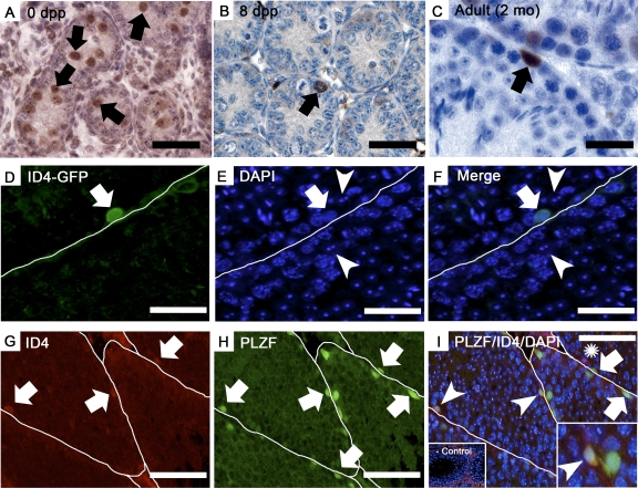
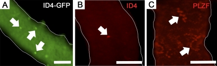
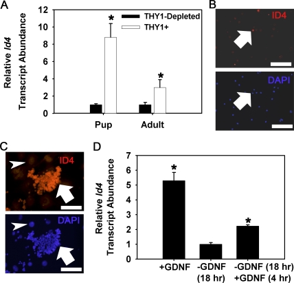
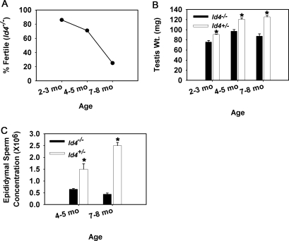
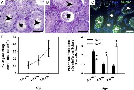
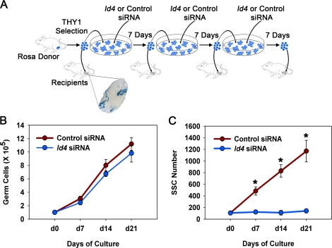
Continual spermatogenesis at a quantitatively normal level is required to sustain male fertility. The foundation of this process relies on maintenance of an undifferentiated spermatogonial population consisting of spermatogonial stem cells (SSCs) that self-renew as well as transient amplifying progenitors produced by differentiation. In mammals, type A(single) spermatogonia form the SSC population, but molecular markers distinguishing these from differentiating progenitors are undefined and knowledge of mechanisms regulating their functions is limited. We show that in the mouse male germline the transcriptional repressor ID4 is expressed by a subpopulation of undifferentiated spermatogonia and selectively marks A(single) spermatogonia. In addition, we found that ID4 expression is up-regulated in isolated SSC-enriched fractions by stimulation from GDNF, a key growth factor driving self-renewal. In mice lacking ID4 expression, quantitatively normal spermatogenesis was found to be impaired due to progressive loss of the undifferentiated spermatogonial population during adulthood. Moreover, reduction of ID4 expression by small interfering RNA treatment abolished the ability of wild-type SSCs to expand in vitro during long-term culture without affecting their survival. Collectively, these results indicate that ID4 is a distinguishing marker of SSCs in the mammalian germline and plays an important role in the regulation of self-renewal.


Figures

FIG. 1
FIG. 1
Expression of ID4 in the undifferentiated spermatogonial population. AC) Immunohistochemistry staining for ID4 expression in cross-sections of testes from mice at the neonatal age of 0 dpp (A), prepubertal age of 8 dpp (B), and adulthood at 2 mo of age (C). DF) Immunofluorescent staining for GFP expression in a testis cross-section from an adult (2 mo of age) homozygous Id4(−)/Gfp(+) knock-in mouse. Green fluorescence indicates GFP(+) expression, which was observed by individual spermatogonia only (arrow) and not by Sertoli cells (arrowhead). DAPI was used to stain all the cell nuclei; white lines mark the basement membrane of adjacent seminiferous tubules. GI) Immunofluorescent staining for ID4 and PLZF expression in cross-sections of testes from adult mice (4 mo of age). ID4 staining (G, arrows) is observed in only a few spermatogonia. In contrast, PLZF staining (H, arrows) is observed in many spermatogonia within seminiferous tubules. In the merged image (I), three different types of spermatogonia are observed, including those that stain for expression of the undifferentiated spermatogonial marker PLZF (arrows), germ cells expressing both PLZF and ID4 (arrowheads), and germ cells expressing ID4 only (asterisks). The left inset is the negative control with normal IgG in place of primary antibodies; note the nonspecific red fluorescence of interstitial cells that can also be observed in cross-sections stained for expression of ID4. The right inset is a magnified view of spermatogonia stained for expression of both PLZF and ID4 (arrowhead). White lines mark the boundaries of seminiferous tubules. Bars are 50 μm for AF and 200 μm for GI.
FIG. 2
FIG. 2
Whole-mount imaging of ID4 expressing cells in mouse seminiferous tubules. A) Representative image of seminiferous tubules from adult homozygous Id4(−)/Gfp(+) knock-in mice. Only single GFP(+) cells were observed (arrows). B) Representative image of immunofluorescent staining (red fluorescence) for expression of ID4 in seminiferous tubules from adult wild-type mice. Only single ID4(+) cells were observed (arrow). C) Representative image of immunofluorescent staining (red fluorescence) for expression of the undifferentiated spermatogonial marker PLZF in seminiferous tubules from adult wild-type mice. In contrast to the staining of ID4 expression in single cells only, PLZF staining is observed in cohorts of spermatogonia (arrows). Bars are 100 μm for A and 50 μm for B and C.
FIG. 3
FIG. 3
Expression of ID4 in the SSC-containing THY1(+) germ cell fraction. A) Quantitative real-time PCR analyses for Id4 gene expression in the THY1(+) germ cell fraction compared to the THY1-depleted testis cell population of testes from prepubertal pup (8 dpp) and adult (3 mo of age) mice. Relative Id4 transcript abundance was calculated by normalization to the constitutively expressed gene Rps2. Data are mean ± SEM for three different MACS-isolated cell populations. *Denotes significant difference at P < 0.05. B) Representative images of immunocytochemical staining for ID4-expressing cells in the MACS-isolated THY1(+) germ cell fraction from prepubertal mice at 8 dpp that contain the entire array of spermatogonia found in the male germline. ID4(+) cells (red fluorescence, arrow) were found to comprise only ∼11% of the cell population. DAPI (blue fluorescence) was used to stain all the cell nuclei. Bars = 100 μm. C) Immunocytochemical staining for ID4 expression in cultured THY1(+) germ cell clumps. Nuclear staining was observed in germ cell clumps (red fluorescence, arrow) but not feeder cell monolayers (arrowhead). DAPI (blue fluorescence) was used to stain all the cell nuclei. Bars are 100 μm. D) Quantitative real-time PCR analyses for GDNF-regulation of Id4 gene expression in cultured THY1(+) germ cells. Germ cell clumps continually cultured with GDNF supplementation (+GDNF) were subjected to 18 h withdrawal of GDNF (−GDNF 18 h) followed by replacement of GDNF for 4 h (+GDNF 4 hr). Relative Id4 transcript abundance was determined by normalization to the constitutively expressed gene Rps2. Data are mean ± SEM for three different THY1(+) germ cell cultures and expressed as fold-difference from the −GDNF treatment. *Denotes significant difference (P < 0.05) from −GDNF treatment.
FIG. 4
FIG. 4
Reproductive phenotype of adult ID4-deficient male mice. A) Fertility assessment of ID4-deficient male mice (Id4−/−) at the developmental stages of young adult (2–3 mo of age), mature adult (4–5 mo of age), and aged (7–8 mo of age). Fertility was assessed based on whether a male sired pups when exposed to two pubertal wild-type female mice. Data are derived from seven Id4−/− male mice at each age point. B) Comparison of testes weights, a measure of normal spermatogenesis, between ID4-deficient mice (Id4−/−) and control mice with sufficient expression of ID4 (Id4+/−) at developmental stages of young adult (2–3 mo of age), mature adult (4–5 mo of age), and aged (7–8 mo of age). Data are mean ± SEM for three different mice of both genotypes at each age point. *Denotes significant difference at P < 0.05. C) Comparison of epididymal sperm concentration between Id4−/− and control Id4+/− male mice at the developmental stages of mature adult (4–5 mo of age) and aged (7–8 mo of age), periods when fertility defects are prominent. Data are mean ± SEM for three different mice of both genotypes at each age point. *Denotes significant difference at P < 0.05.
FIG. 5
FIG. 5
Effects of ID4 loss-of-function on spermatogenesis in testes of adult mice. A and B) Representative images of a cross-section from testes of Id4−/− mice. Seminiferous tubules with depletion of the germ cell population, a Sertoli cell-only phenotype (stars), and qualitatively normal spermatogenesis (arrowheads) were observed. C) Representative image of immunofluorescent staining of a testis cross-section from an adult Id4−/− mouse for expression of the general germ cell marker GCNA1 (green fluorescence). Seminiferous tubules completely lacking germ cells (stars), confirming a Sertoli cell-only phenotype, are observed along with tubules containing germ cells (arrowheads). DAPI (blue fluorescence) was used to stain all the cell nuclei. D) Assessment of the percentage of seminiferous tubules containing a degenerating phenotype and lacking active spermatogenesis in cross-sections of testes from Id4−/− mice at the developmental stages of young adult (2–3 mo of age), mature adult (4–5 mo of age), and aged (7–8 mo of age). Data are mean ± SEM for three different mice at each age point. E) Comparison of the number of undifferentiated spermatogonia in cross-sections of seminiferous tubules from testes of Id4−/− and control Id4+/− mice at young adult (2–3 mo of age), mature adult (4–5 mo of age), and aged (7–8 mo of age) developmental stages. Undifferentiated spermatogonia were identified based on immunohistochemical staining for expression of the marker PLZF. Data are mean ± SEM for three different mice and 75 round seminiferous tubules of both genotypes at each age point. *Denotes significant difference at P < 0.05.
FIG. 6
FIG. 6
Effects of impairing ID4 expression on GDNF-induced self-renewal of SSCs from wild-type mice. A) Experimental strategy for examining the effects of impairing ID4 expression by siRNA treatment on expansion of SSCs in cultures of THY1(+) germ cells supplemented with GDNF over a 21-day period, which constitutes greater than three self-renewal cycles in vitro. THY1(+) germ cells from ROSA donor mice (6–8 dpp) that express a LacZ marker transgene in all the germ cells were treated with nontargeting control or Id4-specific siRNA and maintained in serum-free conditions with GDNF and FGF2 supplementation for 7 days. Single cell suspensions were then created, and a portion of the cells were transplanted into testes of recipient mice to determine SSC content based on the ability to reestablish colonies of spermatogenesis. The remaining cells were again treated with nontargeting control or Id4-specific siRNA and cultured another 7 days before collection and transplantation or retreatment with siRNA. This protocol was conducted three times over the 21-day culture period to track SSC expansion. B) Assessment of overall germ cell expansion in cultures of THY1(+) germ cells treated with nontargeting control or Id4-specific siRNA over a 21-day period. Data are mean ± SEM for three different THY1(+) germ cell cultures. *Denotes significant difference at P < 0.05. C) Assessment of SSC expansion in cultures of THY1(+) germ cells treated with nontargeting control or Id4-specific siRNA over a 21-day period. SSC numbers were determined based on donor-derived colonies of spermatogenesis following transplantation into recipient testes. Data are mean ± SEM for three different THY1(+) germ cell cultures. *Denotes significant difference at P < 0.01. Note that the error bars do not extend beyond the circles for Id4 siRNA treatment.

Comment in

  • The elusive spermatogonial stem cell marker?
    Hermann BP, Phillips BT, Orwig KE. Hermann BP, et al. Biol Reprod. 2011 Aug;85(2):221-3. doi: 10.1095/biolreprod.111.093146. Epub 2011 May 12. Biol Reprod. 2011. PMID: 21565995 Free PMC article.

References

    1. Oatley JM, Brinster RL. Regulation of spermatogonial stem cell self-renewal in mammals. Ann Rev Cell Dev Biol 2008; 24: 263 286. - PMC - PubMed
    1. de Rooij DG, Russell LD. All you wanted to know about spermatogonia but were afraid to ask. J Androl 2000; 21: 776 798. - PubMed
    1. Huckins C. The spermatogonial stem cell population in adult rats. I. Their morphology, proliferation and maturation. Anat Rec 1971; 169: 533 557. - PubMed
    1. Oakberg EF. Spermatogonial stem-cell renewal in the mouse. Anat Rec 1971; 169: 515 531. - PubMed
    1. Nakagawa T, Sharma M, Nabeshima Y, Braun RE, Yoshida S. Functional hierarchy and reversibility within the murine spermatogenic stem cell compartment. Science 2010; 328: 62 67. - PMC - PubMed

Publication types