. 2011 May;67(Pt 5):440-6.
doi: 10.1107/S0907444911007815. Epub 2011 Apr 13.

X-ray crystal structure and small-angle X-ray scattering of sheep liver sorbitol dehydrogenase

Affiliations

X-ray crystal structure and small-angle X-ray scattering of sheep liver sorbitol dehydrogenase

Hemant Yennawar et al. Acta Crystallogr D Biol Crystallogr. 2011 May.

Abstract

The X-ray crystal structure of sheep liver sorbitol dehydrogenase (slSDH) has been determined using the crystal structure of human sorbitol dehydrogenase (hSDH) as a molecular-replacement model. slSDH crystallized in space group I222 with one monomer in the asymmetric unit. A conserved tetramer that superposes well with that seen in hSDH (despite belonging to a different space group) and obeying the 222 crystal symmetry is seen in slSDH. An acetate molecule is bound in the active site, coordinating to the active-site zinc through a water molecule. Glycerol, a substrate of slSDH, also occupies the substrate-binding pocket together with the acetate designed by nature to fit large polyol substrates. The substrate-binding pocket is seen to be in close proximity to the tetramer interface, which explains the need for the structural integrity of the tetramer for enzyme activity. Small-angle X-ray scattering was also used to identify the quaternary structure of the tetramer of slSDH in solution.


Figures

Figure 1
Figure 1
Stereoview of the active site with bound acetate and glycerol molecules. Glycerol is a substrate of SDH. The electron density shown is an OMIT map excluding the zinc, glycerol and acetate and contoured at the 1.0σ level. Zinc-coordination and hydrogen-bond interactions are marked as black dotted lines (distances less than 3.2 Å). The present structure lacks the NAD cofactor in the active site.
Figure 2
Figure 2
A model of NAD and sorbitol binding as guided by the present and human SDH structures.
Figure 3
Figure 3
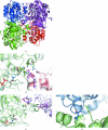
A number of van der Waals contacts and hydrogen bonds stabilize the dimer and tetramer interfaces. Tetramer formation is crucial to strengthen the substrate-binding pockets, shown here occupied by a model of sorbitol (SOR1419) in cyan. A stick model of the NADH cofactor is shown in cyan. (a) The tetramer as seen in the crystal packing of human and sheep liver SDH crystal structures. (b) Interactions seen within a dimer; each monomer forms crucial hydrogen bonds to complete the substrate-binding pocket of the other monomer. (cd) Interactions seen across dimers.
Figure 4
Figure 4
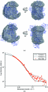
(a) SAXS envelopes superimposed on two candidate tetramers derived from crystal lattice symmetry. Tetramer 1 (top) is structurally analogous to the asymmetric unit of hSDH and is reasonably well contained by the experimental envelope. Tetramer 2 (bottom) is less compact and is not well contained by the envelope. (b) Computed versus experimental scattering for tetramer models. Tetramer 1 most closely matches the experimental data in the region q < 0.1 Å.

Similar articles

Cited by

References

    1. Adams, P. D. et al. (2010). Acta Cryst. D66, 213–221. - PubMed
    1. Banfield, M. J., Salvucci, M. E., Baker, E. N. & Smith, C. A. (2001). J. Mol. Biol. 306, 239–250. - PubMed
    1. Emsley, P. & Cowtan, K. (2004). Acta Cryst. D60, 2126–2132. - PubMed
    1. Engh, R. A. & Huber, R. (1991). Acta Cryst. A47, 392–400.
    1. Franke, D. & Svergun, D. I. (2009). J. Appl. Cryst. 42, 342–346. - PMC - PubMed

Publication types

Substances

Associated data