. 2011 May;67(Pt 5):455-62.
doi: 10.1107/S0907444911008560. Epub 2011 Apr 7.

Structure of a two-CAP-domain protein from the human hookworm parasite Necator americanus

Affiliations

Structure of a two-CAP-domain protein from the human hookworm parasite Necator americanus

Oluwatoyin A Asojo. Acta Crystallogr D Biol Crystallogr. 2011 May.

Abstract

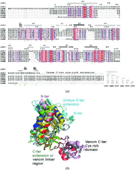
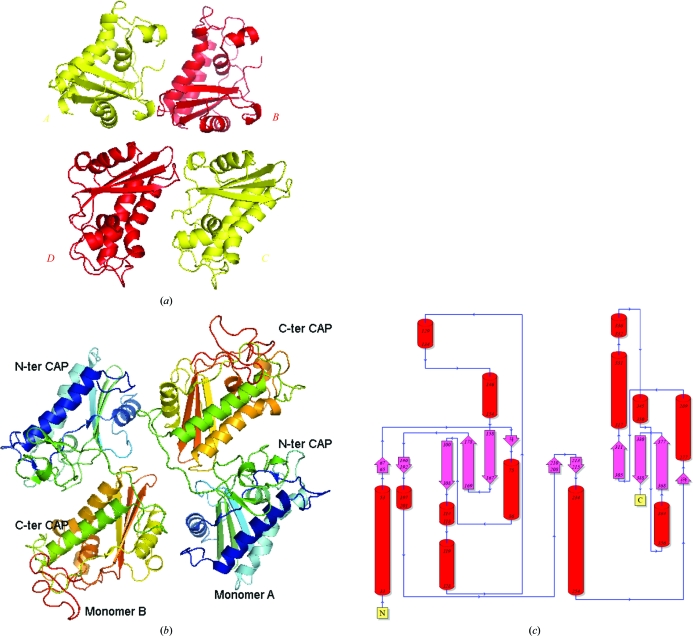
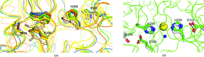
Major proteins secreted by the infective larval stage hookworms upon host entry include Ancylostoma secreted proteins (ASPs), which are characterized by one or two CAP (cysteine-rich secretory protein/antigen 5/pathogenesis related-1) domains. The CAP domain has been reported in diverse phylogenetically unrelated proteins, but has no confirmed function. The first structure of a two-CAP-domain protein, Na-ASP-1, from the major human hookworm parasite Necator americanus was refined to a resolution limit of 2.2 Å. The structure was solved by molecular replacement (MR) using Na-ASP-2, a one-CAP-domain ASP, as the search model. The correct MR solution could only be obtained by truncating the polyalanine model of Na-ASP-2 and removing several loops. The structure reveals two CAP domains linked by an extended loop. Overall, the carboxyl-terminal CAP domain is more similar to Na-ASP-2 than to the amino-terminal CAP domain. A large central cavity extends from the amino-terminal CAP domain to the carboxyl-terminal CAP domain, encompassing the putative CAP-binding cavity. The putative CAP-binding cavity is a characteristic cavity in the carboxyl-terminal CAP domain that contains a His and Glu pair. These residues are conserved in all single-CAP-domain proteins, but are absent in the amino-terminal CAP domain. The conserved His residues are oriented such that they appear to be capable of directly coordinating a zinc ion as observed for CAP proteins from reptile venoms. This first structure of a two-CAP-domain ASP can serve as a template for homology modeling of other two-CAP-domain proteins.


Figures

Figure 1
Figure 1
Structural elements of representative CAPs. (a) The conserved secondary-structure elements of the CAP core are based on Na-ASP-2 (PDB entry 1u53) and the CRISPs (PDB entries 1rc9, 1xx5 and 1wvr). This figure was generated with ESPript (Gouet et al., 2003 ▶). Ves v 5 from yellow jacket (PDB entry 1qnx) has an amino-terminal extension, while both Na-ASP-2 and the CRISPs have carboxyl-terminal extensions. Tomato P14a (PDB entry 1cfe) and human Golgi-associated plant pathogenesis-related protein 1 (GAPR-1; PDB entry 1smb) do not have any extensions. The linker loops of the CRISPs share common sequence features with the carboxyl-terminal s extension of 1u53. (b) Structural alignment of representative CAP proteins; the PDB codes are Na-ASP-2, 1u53 (green); GAPR-1, 1smb (yellow); PI14, 1cfe (blue); snake-venom CRISPs, 1xx5 (red), 1wvr (pink) and 1rc9 (gray). All core helices and strands overlay well from the mammalian (1smb), plant (1cfe), snake-venom CRISP (1xx5, 1wvr and 1rc9) and nematode (1u53) SCP domains. Snake-venom CRISPs have a unique C-terminal extension and 1qnx has a unique N-terminal extension. Despite the varied sequence homology, the three-dimensional structures of representative CAP proteins reveal significant structural homology in a conserved α/β-barrel core.
Figure 2
Figure 2
Structure of Na-ASP-1. (a) A ribbon diagram of the molecular-replacement solution shows the proximity of the four CAP domains and illuminates the difficulty in assigning the connectivity of the domains. (b) Ribbon diagram of crystallographic dimer of Na-ASP-1 rainbow colored from blue (N-­terminus) to red (C-terminus). (c) Topology plot of Na-ASP-1 generated with PDBSum (http://www.ebi.ac.uk/thornton-srv/databases/pdbsum/).
Figure 3
Figure 3
Comparison of Na-ASP-1 with selected representative two-CAP-domain proteins. Sequence alignment of selected two-CAP-domain proteins reveals the sequence conservation across both domains. The highest variability is in the signal peptide and loop regions. This figure was generated with ESPript. The different secondary-structure elements shown are α-helices as large squiggles labelled α, 310-helices as small squiggles labelled η, β-strands as arrows labelled β and β-turns labelled TT. Identical residues are shown on a red background, conserved residues are shown in red and conserved regions are shown in blue boxes. Na-ASP-2 (AAP41952.1) is included as a representative one-CAP-domain ASP. Aligned proteins are Na-ASP-1, N. americanus ASP-1 (AAD13340.1); Ac-ASP-1, Ancylostoma caninum ASP-1 (AAD318391); Ad-ASP-1, A. duodenale ASP-1 (AAD13339.1); Acy-ASP-1, A. ceylanicum ASP-1 (AAN11402.1); Cre-VAP-1, Caenorhabditis remanei two-domain activation-associated secreted protein (XP_003106746.1); Cbr-VAP-1, C. briggsae VAP-1 (CAP3467.2); Ce-VAP-1, C. elegans VAP-1 (NM_001029382.1); Cre-SCL22, C. remanei two-domain activation-associated secreted protein (XP_003109641.1); Cp-ASP1b, Cooperia punctata two-domain activation-associated secreted protein-like (AAK35199.1); Acy-ASP-5, A. ceylanicum ASP-5 (ABB53347.1); Oo-VAP-4, Ostertagia ostertagi VAP-4 (CA000417); Hc-ASP-1, Haemonchus contortus putative secretory protein precursor (AAC03562). GenBank accession numbers are given in parentheses. The sequence identity of each protein is also listed.
Figure 4
Figure 4
Surface plots of representative CAP structures in green reveal a conserved putative binding cavity. The plots of (a) Na-ASP-1, (b) Na-ASP-2 (1u53), (c) Ves v 5 (1qnx), (d) snake-venom CRISP (1xx5) and (e) GAPR-1 (1smb) are shown the same orientation and the location of the two conserved Glu and His residues are shown in red. The amino-terminal CAP cavity of Na-ASP-1 is colored blue. The extension of the cavity through the entire monomer of Na-ASP-1 is reminiscent of the CRISPs in which the cavity extends from the amino-terminal CAP to the carboxyl-terminal cysteine-rich domain.
Figure 5
Figure 5
Superposed conserved central cavity of CAPs. (a) The superposed cavities reveal that key residues corresponding to His295, Glu306, Glu332, His358 and Ser296 superimpose well in representative CAP structures. CAP structures are colored as follows: Na-ASP-1, green; Na-ASP-2, orange; Ves v 5, yellow; GAPR-1, brown; snake-venom CRISP (1xx5), blue. (b) These same residues superpose in Na-ASP-1 (green), with the cobra CRISP natrin (3mz8) in complex with Zn2+(gray). Zn2+ is shown in yellow and a water molecule that is coordinated to the Zn2+ is shown in blue.
Figure 6
Figure 6
Domain association of covalently linked CAP domains of Na-ASP-1. The C-terminal CAP domain of Na-ASP-1 (shown in aquamarine) is blocked by the N-terminal CAP domain (gray and pink) and the linker loop (yellow). Also shown are the first 96 residues of the N-terminal CAP domain (pink) that when truncated expose more of the C-terminal CAP domain and result in better antibody binding.

References

    1. Asojo, O. A., Goud, G., Dhar, K., Loukas, A., Zhan, B., Deumic, V., Liu, S., Borgstahl, G. E. & Hotez, P. J. (2005). J. Mol. Biol. 346, 801–814. - PubMed
    1. Asojo, O. A., Loukas, A., Inan, M., Barent, R., Huang, J., Plantz, B., Swanson, A., Gouthro, M., Meagher, M. M. & Hotez, P. J. (2005). Acta Cryst. F61, 391–394. - PMC - PubMed
    1. Asojo, O. A., Loukas, A., Inan, M., Barent, R., Huang, J., Plantz, B., Swanson, A., Gouthro, M., Meagher, M. M. & Hotez, P. J. (2010). Acta Cryst. F66, 1549. - PMC - PubMed
    1. Bethony, J., Loukas, A., Smout, M., Brooker, S., Mendez, S., Plieskatt, J., Goud, G., Bottazzi, M. E., Zhan, B., Wang, Y., Williamson, A., Lustigman, S., Correa-Oliveira, R., Xiao, S. & Hotez, P. J. (2005). FASEB J. 19, 1743–1745. - PubMed
    1. Bethony, J. M., Simon, G., Diemert, D. J., Parenti, D., Desrosiers, A., Schuck, S., Fujiwara, R., Santiago, H. & Hotez, P. J. (2008). Vaccine, 26, 2408–2417. - PubMed

Publication types

Substances

Associated data