. 2011 Jun 1;10(11):1845-52.
doi: 10.4161/cc.10.11.15777. Epub 2011 Jun 1.

MicroRNA signatures differentiate melanoma subtypes

Affiliations

MicroRNA signatures differentiate melanoma subtypes

Elcie Chan et al. Cell Cycle. .

Abstract

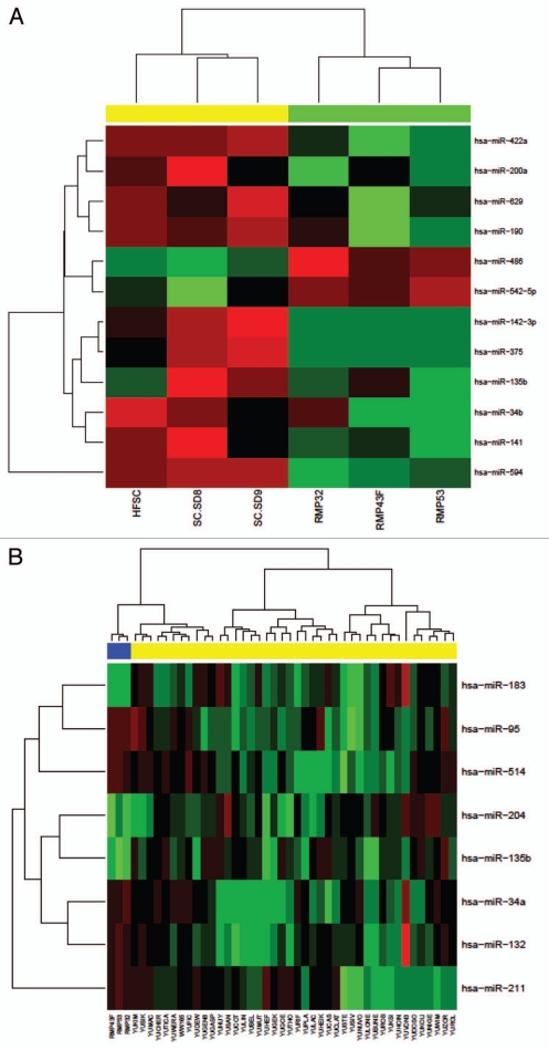
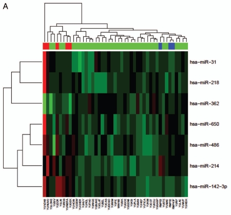
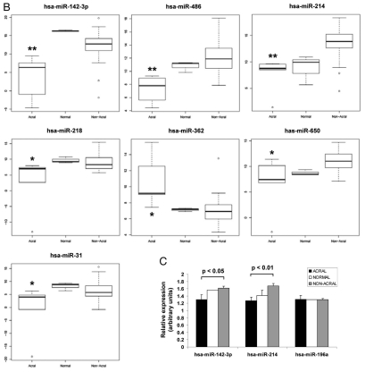
Melanoma is an aggressive cancer that is highly resistance to therapies once metastasized. We studied microRNA (miRNA) expression in clinical melanoma subtypes and evaluated different miRNA signatures in the background of gain of function somatic and inherited mutations associated with melanoma. Total RNA from 42 patient derived primary melanoma cell lines and three independent normal primary melanocyte cell cultures was evaluated by miRNA array. MiRNA expression was then analyzed comparing subtypes and additional clinicopathologic criteria including somatic mutations. The prevalence and association of an inherited variant in a miRNA binding site in the 3'UTR of the KRAS oncogene, referred to as the KRAS-variant, was also evaluated. We show that seven miRNAs, miR-142-3p, miR-486, miR-214, miR-218, miR-362, miR-650 and miR-31, were significantly correlated with acral as compared to non-acral melanomas (p < 0.04). In addition, we discovered that the KRAS-variant was enriched in non-acral melanoma (25%), and that miR-137 under expression was significantly associated with melanomas with the KRAS-variant. Our findings indicate that miRNAs are differentially expressed in melanoma subtypes and that their misregulation can be impacted by inherited gene variants, supporting the hypothesis that miRNA misregulation reflects biological differences in melanoma.


Figures

Figure 1
Figure 1
Melanoma microRNA expression profiles. (A) Newborn foreskin melanocyte and normal adult melanocyte samples are arranged in columns on the horizontal axis with newborn melanocytes represented in three yellow columns on the left, and adult melanocytes in the three green columns on the right. (B) The top most differentially expressed miRNAs between normal melanocytes and melanoma cell lines. The miRNA profiles of 42 melanoma samples plus three control samples were assayed by Taqman miRNA expression array. Cell lines are arranged in columns on the horizontal axis with normal control melanocytes represented in blue.
Figure 2
Figure 2
Acral melanomas exhibit significantly differential expression of seven miRNAs relative to other melanomas. (A) Clustered expression of miR-31, miR-218, miR-362, miR-650, miR-486, miR-214 and miR-142-3p in melanomas. Cell lines are arranged in columns on the horizontal axis with acral melanomas represented in red and normal melanocytes in blue. (B) Box-whisker plots of normalized miRNA expression in acral (n = 5), non-acral (n = 37) and normal melanocytes (n = 3). The vertical axis represents average comparative CT values, where higher values correspond to low relative expression. The top and bottom lines of the boxes represent the upper and lower quartile respectively, the center line represents the median, and the whiskers show the max/min range. Multiple testing corrected p-values for acral versus nonacral melanomas are shown, **p < 0.01, *p < 0.04. (C) Quantitative RT-PCR secondary validation of miRNA expression. The expression of miR-142-3p and miR-214, in addition to miR-196a as a control was validated in a random subset of normal (n = 2), acral (n = 4) and non-acral melanomas (n = 6). All expression values (arbitrary units) were standardised to RNU44 and the standard error is shown. The p-value between acral and non-acral melanomas was calculated by unpaired t-test.
Figure 2
Figure 2
Acral melanomas exhibit significantly differential expression of seven miRNAs relative to other melanomas. (A) Clustered expression of miR-31, miR-218, miR-362, miR-650, miR-486, miR-214 and miR-142-3p in melanomas. Cell lines are arranged in columns on the horizontal axis with acral melanomas represented in red and normal melanocytes in blue. (B) Box-whisker plots of normalized miRNA expression in acral (n = 5), non-acral (n = 37) and normal melanocytes (n = 3). The vertical axis represents average comparative CT values, where higher values correspond to low relative expression. The top and bottom lines of the boxes represent the upper and lower quartile respectively, the center line represents the median, and the whiskers show the max/min range. Multiple testing corrected p-values for acral versus nonacral melanomas are shown, **p < 0.01, *p < 0.04. (C) Quantitative RT-PCR secondary validation of miRNA expression. The expression of miR-142-3p and miR-214, in addition to miR-196a as a control was validated in a random subset of normal (n = 2), acral (n = 4) and non-acral melanomas (n = 6). All expression values (arbitrary units) were standardised to RNU44 and the standard error is shown. The p-value between acral and non-acral melanomas was calculated by unpaired t-test.
Figure 3
Figure 3
MiR-137 differentially associates with the KRAS-variant in melanomas. (A) Box-whisker plot of miR-137 expression in melanoma samples with (MUT, n = 8) and without (WT, n = 29) the KRAS-variant, p < 0.011 after multiple testing correction. The vertical axis represents average comparative CT values, where higher values correspond to low relative expression. The top and bottom lines of the boxes represent the upper and lower quartile respectively, the center line represents the median, and the whiskers show the max/min range. (B) Microarray gene expression of miR-137 associated genes, *p < 0.05, -1 transcript variant 1, -2 transcript variant 2.

Comment in

Similar articles

Cited by

References

    1. Liu V, Mihm MC. Pathology of malignant melanoma. Surg Clin North Am. 2003;83:31–60. - PubMed
    1. Esquela-Kerscher A, Slack FJ. Oncomirs—microRNAs with a role in cancer. Nat Rev Cancer. 2006;6:259–269. - PubMed
    1. Babar IA, Slack FJ, Weidhaas JB. miRNA modulation of the cellular stress response. Future Oncol. 2008;4:289–298. - PubMed
    1. Bartel DP. MicroRNAs: Genomics, biogenesis, mechanism and function. Cell. 2004;116:281–297. - PubMed
    1. Lu J, Getz G, Miska EA, Alvarez-Saavedra E, Lamb J, Peck D, et al. MicroRNA expression profiles classify human cancers. Nature. 2005;435:834–838. - PubMed

Publication types