. 2011 Oct;80(7):719-30.
doi: 10.1038/ki.2011.122. Epub 2011 May 4.

The inducible deletion of Drosha and microRNAs in mature podocytes results in a collapsing glomerulopathy

Affiliations

The inducible deletion of Drosha and microRNAs in mature podocytes results in a collapsing glomerulopathy

Olga Zhdanova et al. Kidney Int. 2011 Oct.

Abstract

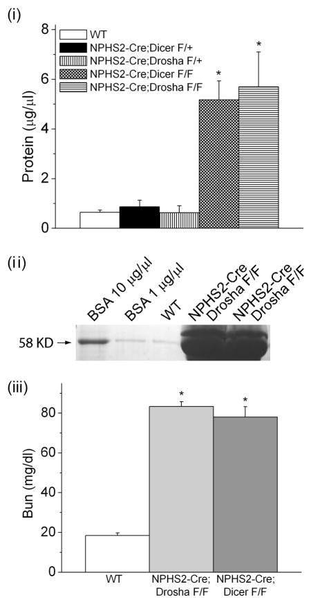
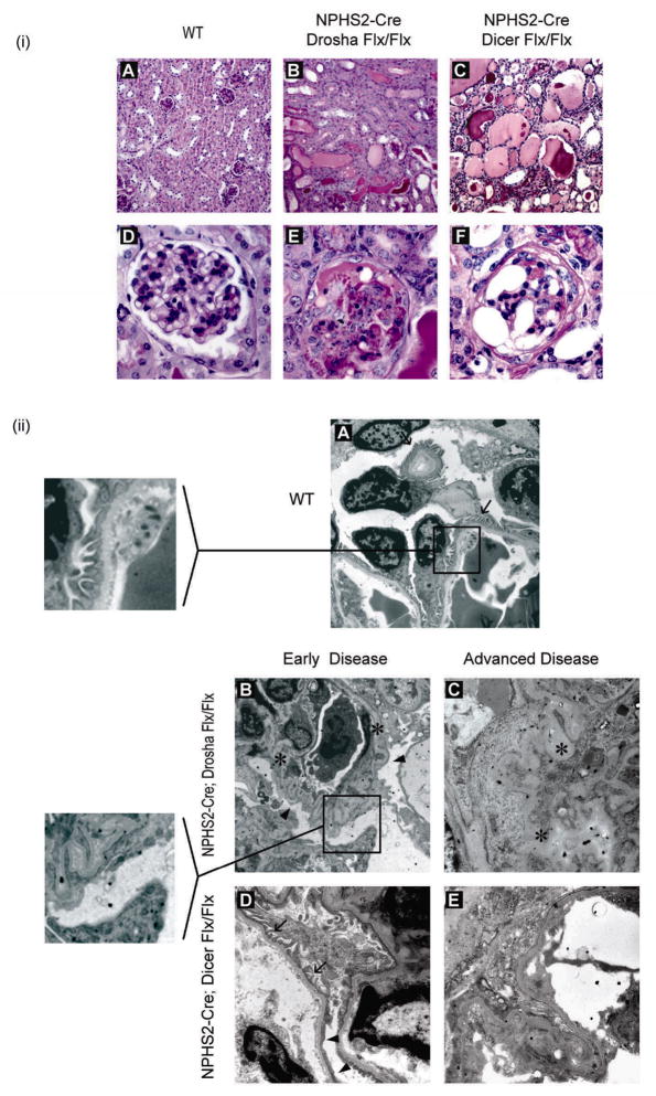
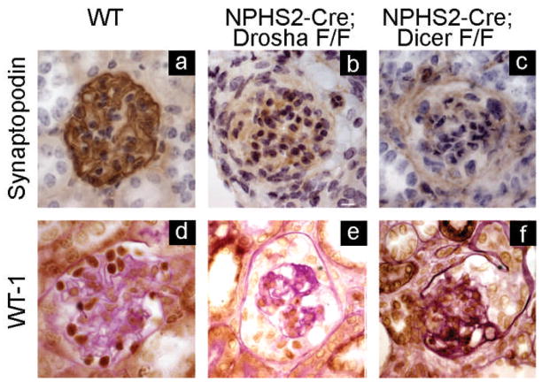
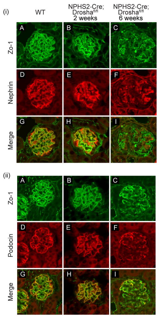
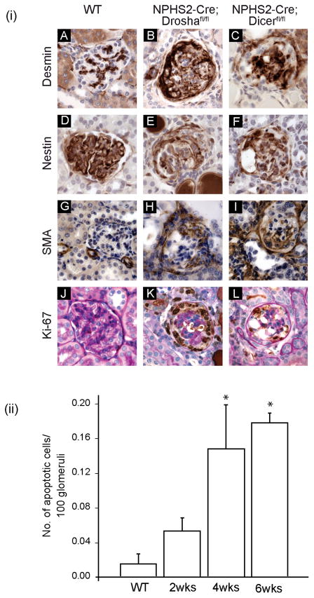
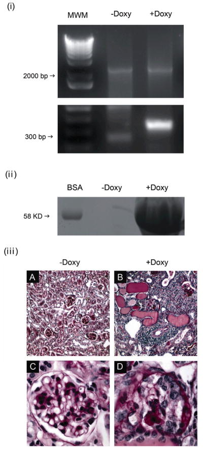
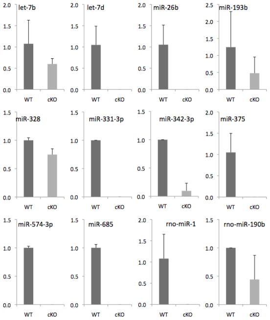
Micro-RNAs (miRNAs) are short (average 22 nucleotides) noncoding regulatory RNAs that inhibit gene expression by targeting complementary 3'-untranslated regions of protein-encoding mRNAs for translational repression or degradation. miRNAs play key roles in both the function and differentiation of many cell types. Drosha and Dicer, two RNAase III enzymes, function in a stepwise manner to generate a mature miRNA. Previous studies have shown that podocyte-specific deletion of Dicer during development results in proteinuric renal disease and collapsing glomerulopathy (CG); however, Dicer has functions other than the generation of miRNAs. Here we found that the podocyte-specific deletion of Drosha results in a similar phenotype to Dicer mutants, confirming that the Dicer mutant phenotype is due to the loss of miRNAs. Moreover, the inducible deletion of Drosha in 2- to 3-month-old mice (Tet-On system) resulted in CG. Thus, continuous generation of miRNAs are required for the normal function of mature podocytes and their loss leads to CG. Identifying these miRNAs may provide new insight into disease pathogenesis and novel therapeutic targets in various podocytopathies.


Conflict of interest statement

Disclosure: All the authors declared no competing interests.

Figures

Figure 1
Figure 1. NPHS2-Cre; Droshafl/fl develop proteinuria and renal failure
(i) Protein concentration in urine obtained from WT (NPHS2-Cre; Drosha+/+), heterozygous (NPHS2-Cre; Droshafl/+), and homozygous Drosha (NPHS2-Cre; Droshafl/fl) and Dicer (NPHS2-Cre; Dicerfl/fl ) mutants determined by Bradford assay (n=5 mice in each group). Differences in protein concentrations were statistically significant for homozygous Drosha and Dicer mice when compared with WT and homozygous animals (* P<0.05). (ii) 10 ul of urine from 4 week old WT and homozygous Drosha mutants were separated by SDS polyacrylamide gel electrophoreses (10%) followed by coumassie staining. Bovine serum albumin (BSA) was run as a control. (iii) Bun concentration was determined in 4 week old WT and homozygous Drosha and Dicer mutants (n= 5 mice in each group). Bun values were statistically significant in homozygous Drosha and Dicer mice when compared to WT mice (* P<0.05).
Figure 2
Figure 2. NPHS2-Cre; Droshafl/fl develop collapsing glomerulopathy with focal glomerulosclerosis with pseudocrescent and microcystic dilatation of tubules
(i) Histologic examination (PAS stain) of kidneys from WT (NPHS2-Cre; Drosha+/+) and homozygous Drosha (NPHS2-Cre; Droshafl/fl) and Dicer (NPHS2-Cre; Dicerfl/fl ) mutants at 4–5 weeks of age. Upper panel, 20X magnification (A–C); lower panel, 40X magnification (D–F). (ii) Electron microscopy of kidneys from WT (A) or homozygous Drosha and Dicer mutants at 2 weeks (early disease-B, D) or 4–5 weeks (advanced disease-C,E) of age. Arrows indicate normal podocyte foot processes. Arrowheads indicate effaced podocyte foot processes. Asterisks indicate collapsed glomerular basement membrane. Insert in panel A shows normal podocyte foot processes. Insert in Panel B shows effaced podocyte foot processes.
Figure 3
Figure 3. Podocytes from Drosha and Dicer mutants have decreased expression of synaptopodin and WT-1
Immunohistochemistry of histologic sections from 4–5 week old WT, Drosha, and Dicer mutants immunostained with antibodies to synaptopodin and WT-1. Podocyte expression of synaptopodin and WT are decreased in both Drosha and Dicer mutants.
Figure 4
Figure 4. Expression of podocin and nephrin in WT and Drosha mutants
Immunofluorescence of histologic sections from WT and Drosha mutants at 2 and 6 weeks of age stained with antibodies to (i) nephrin (D–F) and (ii) podocin (D–F). Sections were also stained with anit-ZO1 to mark adherent junctions on podocytes (i) A–C and (ii) A–C.
Figure 5
Figure 5. Pseudocrescents stain positive for desmin, nestin, SMA, and Ki-67
(i) Immunohistochemistry of histological sections from 4–5 week old WT (NPHS2-Cre; Drosha+/+), homozygous Drosha (NPHS2-Cre; Droshafl/fl) and Dicer (NPHS2-Cre; Dicerfl/fl ) mutants immunostained with antibodies as indicated. (ii) NPHS2-Cre; Droshafl/fl demonstrate increased apoptosis as CG becomes more advanced. Statistically significant for 4 and 6 week old NPHS2-Cre; Droshafl/fl when compared to WT animals or 2 week old NPHS2-Cre; Droshafl/fl (* P<0.05).
Figure 6
Figure 6. Conditional inducible deletion of Drosha in podocytes in 2 month old animals results in a collapsing glomerulopathy
(i) Glomeruli were isolated from Droshafl/fl;podocin-rtTAtg/+; tetO-Cretg/+ mice that were either administered or not administered doxycycline and deletion of exon 9 was assessed by PCR using primers that flank the loxP site. Deletion of exon 9 results in a product of 334 base pairs, in contrast to the 2.5 Kb product in the wild type gene. 1kB plus Invitrogen was used as molecular weight marker (MWM) (ii) 10 ul of urine from Droshafl/fl;podocin-rtTAtg/+; tetO-Cretg/+ mice that were administered or not administered doxycycline for 2–4 weeks were separated by SDS polyacrylamide gel electrophoreses (10%) followed by coumassie staining. Bovine serum albumin (BSA) was run as a control. (iii) Droshafl/fl;podocin-rtTAtg/+; tetO-Cretg/+ mice were either administered, or not administered doxycycline at 2 months of age and kidneys were analyzed 1 month later. Shown are PAS stains of kidneys sections from (A,C) Dox-minus and (B,D) Dox-plus animals. Figure (A) shows normal glomeruli and unremarkable tubule-interstitum (20X). On higher magnification (C, 40X), glomeruli are normal in size and cellularity and mesangium is normally expanded. Glomerular basement membranes are also unremarkable. No proliferation in the urinary space is noted. In contrast, animals fed doxycycline (B, 20X) had numerous tubular microcysts and a dense interstitial inflammation, which on higher magnification (D, 40X) revealed shrunken glomeruli, with collapsed capillary walls and proliferation of epithelial cells that filled the urinary space.
Figure 7
Figure 7. miRNA expression in glomeruli from Doxycycline-minus and Doxycyline-plus animals
2 month old Droshafl/fl;podocin-rtTAtg/+; tetO-CreTg/+ mice were either administered or not administered doxycycline for 2 weeks and glomeruli were isolated by laser capture microdissection as described in Materials and Methods. RNA was then reversed transcribed and miRNA expression was quantitated using Taqman Low Density Microarrays (Applied Biosystems). Shown are 10 miRNAs that were consistently markedly downregulated in doxycycline treated animals from 3 independent experiments.

Comment in

References

    1. Bartel DP. MicroRNAs: genomics, biogenesis, mechanism, and function. Cell. 2004;116:281–297. - PubMed
    1. Rana TM. Illuminating the silence: understanding the structure and function of small RNAs. Nat Rev Mol Cell Biol. 2007;8:23–36. - PubMed
    1. Filipowicz W, Bhattacharyya SN, Sonenberg N. Mechanisms of post-transcriptional regulation by microRNAs: are the answers in sight? Nat Rev Genet. 2008;9:102–114. - PubMed
    1. Eulalio A, Huntzinger E, Izaurralde E. Getting to the root of miRNA-mediated gene silencing. Cell. 2008;132:9–14. - PubMed
    1. Lee Y, Ahn C, Han J, et al. The nuclear RNase III Drosha initiates microRNA processing. Nature. 2003;425:415–419. - PubMed

Publication types