. 2011 Apr 22;6(4):e19003.
doi: 10.1371/journal.pone.0019003.

Isolation and mutagenesis of a capsule-like complex (CLC) from Francisella tularensis, and contribution of the CLC to F. tularensis virulence in mice

Affiliations

Isolation and mutagenesis of a capsule-like complex (CLC) from Francisella tularensis, and contribution of the CLC to F. tularensis virulence in mice

Aloka B Bandara et al. PLoS One. .

Abstract

Background: Francisella tularensis is a category-A select agent and is responsible for tularemia in humans and animals. The surface components of F. tularensis that contribute to virulence are not well characterized. An electron-dense capsule has been postulated to be present around F. tularensis based primarily on electron microscopy, but this specific antigen has not been isolated or characterized.

Methods and findings: A capsule-like complex (CLC) was effectively extracted from the cell surface of an F. tularensis live vaccine strain (LVS) lacking O-antigen with 0.5% phenol after 10 passages in defined medium broth and growth on defined medium agar for 5 days at 32°C in 7% CO₂. The large molecular size CLC was extracted by enzyme digestion, ethanol precipitation, and ultracentrifugation, and consisted of glucose, galactose, mannose, and Proteinase K-resistant protein. Quantitative reverse transcriptase PCR showed that expression of genes in a putative polysaccharide locus in the LVS genome (FTL_1432 through FTL_1421) was upregulated when CLC expression was enhanced. Open reading frames FTL_1423 and FLT_1422, which have homology to genes encoding for glycosyl transferases, were deleted by allelic exchange, and the resulting mutant after passage in broth (LVSΔ1423/1422_P10) lacked most or all of the CLC, as determined by electron microscopy, and CLC isolation and analysis. Complementation of LVSΔ1423/1422 and subsequent passage in broth restored CLC expression. LVSΔ1423/1422_P10 was attenuated in BALB/c mice inoculated intranasally (IN) and intraperitoneally with greater than 80 times and 270 times the LVS LD₅₀, respectively. Following immunization, mice challenged IN with over 700 times the LD₅₀ of LVS remained healthy and asymptomatic.

Conclusions: Our results indicated that the CLC may be a glycoprotein, FTL_1422 and -FTL_1423 were involved in CLC biosynthesis, the CLC contributed to the virulence of F. tularensis LVS, and a CLC-deficient mutant of LVS can protect mice against challenge with the parent strain.


Conflict of interest statement

Competing Interests: The authors have declared that no competing interests exist.

Figures

Figure 1
Figure 1. Negative stain electron microscopy of the CLC of F. tularensis.
Panels A and E: type B strain LVS_P10 grown at 32°C on Glc-CDMA; different cultures were examined on different days; Panel B: type A strain TI0902, passed and grown to enhance CLC expression as for LVS. In this and some other cases the CLC appeared to aggregate, which also occurred following isolation of the CLC; Panel C: glycosyl transferase mutant LVSΔ1423/1422_P10; Panel D: complemented strain LVSΔ1423/1422[1423/1422+]_P10; Panel F: LVS not passed in defined medium and grown on CDMA at 37°C; only a small amount of CLC is visible (arrow); Panel G: O-antigen mutant WbtIG191V_P17 grown as for LVS_P10; Panel H: O-antigen and CLC double mutant WbtIG191V_P17Δ1423/1422. Strains LVS_P10, WbtIG191V_P17, and type A strain TI0902_P10 have an electron dense layer surrounding their cells. This layer is missing in mutants LVSΔ1423/1422_P10 and WbtIG191V_P17Δ1423/1422, and is restored in complemented strain LVSΔ1423/1422[1423/1422+]_P10]. The bacteria were fixed in glutaraldehyde, and stained with uranyl acetate. Magnification is 20,000 X, and the scale bar is 500 nm.
Figure 2
Figure 2. Polyacrylamide gel electrophoresis of CLC extracts.
CLC extracts at various stages of purity, or F. tularensis LPS as a control, were separated by electrophoresis in a 4–12% separating gel, and the components identified by (A), Coomassie Blue for protein; (B), Stains-All/silver stain for acidic molecules; (C), Western blot with antiserum to LVS whole cells for antigenic components; (D), Pro-Q Emerald stain for carbohydrate. Lanes for panels A-C: M, molecular size standards; 1, LVS LPS (20 µg); 2, crude CLC prior to enzyme digestion (20 µg); 3, CLC extract following enzyme digestion (20 µg); 4, purified CLC (20 µg). Lanes for panel D: M, molecular size standards; 1, crude CLC prior to enzyme digestion (20 µg); 2, CLC extract following enzyme digestion (20 µg); 3, purified CLC (20 µg). The Pro-Q Emerald stain of LPS (not shown here) looks similar to that of the Western blot, and is shown in reference 29.
Figure 3
Figure 3. Trimethylsilyl (TMS) ethers of the methyl glycosides from purified F. tularensis CLC.
TMS derivatives of the glycoses from the CLC were analyzed by GC/MS. All sugars and C18 fatty acids match in both retention time and mass spectrum with known standards.
Figure 4
Figure 4. RT-qPCR of various regions of the putative CLC locus.
LVS was passed in defined medium and grown on defined medium agar at 32°C in CO2 to maximize CLC content, or in defined medium broth at 37°C without preliminary passage to minimize CLC. The RNA was isolated, converted to cDNA, and the cDNA amplified by quantitative real-time PCR. The results are shown as the x-fold change in gene expression using LVS grown in BHIC broth to log phase (minimal CLC expression) as the calibrator and GAPDH as the endogenous control for gene expression. The locus tag of each gene from LVS is shown on the X-axis.
Figure 5
Figure 5. CLC content from LVS_P10, LVSΔ1423/1422_P10, and LVSΔ1423/1422[1423/1422+]_P10.
Lanes: 1, LVS_P10; 2, LVSΔ1423/1422_P10; 3, LVSΔ1423/1422[1423/1422+]_P10. A) Carbohydrate and protein content of extracted CLC; B) Stains All/silver stain of extracted CLC. The reducing carbohydrate content was measured by phenol sulfuric acid assay , and the protein content was measured by BCA assay. The CLC was extracted from the same number of cells of each strain, as described in Materials and Methods.
Figure 6
Figure 6. RT-PCR of the DNA region FTL_1421 from LVSΔ1423/1422.
LVS and LVSΔ1423/1422 were grown on Glc-CDMA for at least 2 days, the RNA isolated, converted to cDNA, and amplified by PCR to determine if a transcript from DNA downstream of the mutation was made. Lanes: M, 1 kb+ DNA molecular size standards; 1, control amplification of FTL_1425-1424 in LVS; 2, amplification of FTL_1425-1424 upstream of the mutation in LVSΔ1423-1422; 3, control amplification of FTL_1425-1424 upstream of the mutation (no bacteria); 4, control amplification of FTL_1421 from LVS; 5, amplification of FTL_1421 immediately downstream of the mutation in LVSΔ1423/1422. The presence of a normal band of about 351 bp from LVSΔ1423/1422 indicated that the mutation had no polar effect on downstream genes.
Figure 7
Figure 7. Bactericidal assay of LVSΔ1423/1422 and control strains in the presence of fresh human serum.
The bacteria were diluted in PBS supplemented with 0.15 mM CaCl2, 0.5 mM MgCl2, and 2%, 4%, 8%, 16%, or 20% fresh, pooled human serum. Aliquots were cultured by viable plate count before and after 60 min. incubation at 37°C. Bacterial strains: LVS, __________; LVSΔ1423/1422, ---▴---; O-antigen mutant WbtIG191V_P17, __ __ __ __.
Figure 8
Figure 8. Intracellular survival of F. tularensis LVSΔ1423/1422 in J774A.1 cells.
The J774A.1 monolayer (approximately 4.5×10⁵ macrophages/well) was infected with approximately 5.5×10⁷ CFU/well of strain LVS (), LVSΔ1423/1422 (▴), or LVSΔ1423/1422[1423/1422+] (X). Intracellular survival of the bacteria was determined at 0, 24, 48, and 72 h post-infection, as described in Materials and Methods. Data are shown on the log scale as the average number of bacteria recovered from dilutions of lysates of J774A.1 cells. The results shown were from two experiments tested in duplicate at each time point. The slopes for intracellular growth of the bacteria as log10 CFU/well between the 24th and the 48th hour for strains LVS, LVSΔ1423/1422, and LVSΔ1423/1422[1423/1422+] were +1.44, +0.73, and +0.97, respectively.
Figure 9
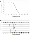
Figure 9. Survival of mice inoculated with F. tularensis LVSΔ1423/1422.
Groups of BALB/c mice were inoculated IN (A) or IP (B), and survival was monitored for six weeks. No mice challenged IN with LVSΔ1423/1422 died during the study. The doses and symbols used for IN inoculations were about 1.2×10⁴ CFU/mouse of LVS (), and about 1.6×10⁴ CFU of LVSΔ1423/1422 (▴). The doses and symbols used for IP inoculations were 262 CFU/mouse of LVS (); 3,375 CFU/mouse of LVS (x); 1,124 CFU/mouse of LVSΔ1423/1422 (▴); 11,136 CFU/mouse of LVSΔ1423/1422 (); 33,408 CFU/mouse of LVSΔ1423/1422 (▪).
Figure 10
Figure 10. Recovery of F. tularensis LVSΔ1423/1422 from the tissues of mice following IN inoculation.
Groups of BALB/c mice were inoculated IN with 7.9×10³ CFU of strain LVS, 5.0×10⁴ CFU of strain LVSΔ1423/1422 (high dose of mutant), or 1.1×10⁴ CFU of strain LVSΔ1423/1422 (low dose of mutant). At 2, 4, or 7 days PI, mice were euthanized. The lungs (A), liver (B), and spleen (C) were aseptically removed, homogenized in PBS, and the CFU/g of tissue determined. The recovery of bacteria from inoculated mice are shown as dark-filled bars (LVS), open bars (high dose of LVSΔ1423/1422), and grey-filled bars (low dose of LVSΔ1423/1422). The mean values of the CFUs of each dose of the mutant were separately compared with LVS. The P values for the differences between the mean values were <0.05 (*) or <0.005 (*).
Figure 11
Figure 11. Protective efficacy of LVSΔ1423/1422 against IN challenge of mice with LVS.
Groups of BALB/c mice were injected with PBS (•), or inoculated with 6.1×10³ CFU/mouse of LVS Δ1423/1422 (). Six weeks PI, the mice were challenged IN with 1.4×10⁵ CFU/mouse of LVS, and the mice were monitored for 4 weeks. No mice died or were symptomatic by 10 days post-challenge. The P value for animal survival after 10 days post-challenge was <0.001.

References

    1. Ellis J, Oyston PC, Green M, Titball RW. Tularemia. Clin Microbiol Rev. 2002;15:631–646. - PMC - PubMed
    1. Dennis DT, Inglesby TV, Henderson DA, Bartlett JG, Ascher MS, et al. Tularemia as a biological weapon: medical and public health management. JAMA. 2001;285:2763–2773. - PubMed
    1. Staples JE, Kubota KA, Chalcraft LG, Mead PS, Petersen JM. Epidemiologic and molecular analysis of human tularemia, United States, 1964-2004. Emerg Infect Dis. 2006;12:1113–1118. - PMC - PubMed
    1. Penn RL. Francisella tularensis (Tularemia). In: Mandell GL, Bennett JE, Dolin R, editors. Mandell, Douglas, and Bennett's Principals and Practice of Infectious Diseases. 6th ed. Philadelphia: Elsevier Churchill Livingstone; 2005. pp. 2674–2685.
    1. Tigertt WD. Soviet viable Pasteurella tularensis vaccines. A review of selected articles. Bacteriol Rev. 1962;26:354–373. - PMC - PubMed

Publication types

MeSH terms