. 2011 Apr 22;6(4):e18969.
doi: 10.1371/journal.pone.0018969.

Variation in array size, monomer composition and expression of the macrosatellite DXZ4

Affiliations

Variation in array size, monomer composition and expression of the macrosatellite DXZ4

Deanna C Tremblay et al. PLoS One. .

Abstract

Macrosatellites are some of the most polymorphic regions of the human genome, yet many remain uncharacterized despite the association of some arrays with disease susceptibility. This study sought to explore the polymorphic nature of the X-linked macrosatellite DXZ4. Four aspects of DXZ4 were explored in detail, including tandem repeat copy number variation, array instability, monomer sequence polymorphism and array expression. DXZ4 arrays contained between 12 and 100 3.0 kb repeat units with an average array containing 57. Monomers were confirmed to be arranged in uninterrupted tandem arrays by restriction digest analysis and extended fiber FISH, and therefore DXZ4 encompasses 36-288 kb of Xq23. Transmission of DXZ4 through three generations in three families displayed a high degree of meiotic instability (8.3%), consistent with other macrosatellite arrays, further highlighting the unstable nature of these sequences in the human genome. Subcloning and sequencing of complete DXZ4 monomers identified numerous single nucleotide polymorphisms and alleles for the three microsatellite repeats located within each monomer. Pairwise comparisons of DXZ4 monomer sequences revealed that repeat units from an array are more similar to one another than those originating from different arrays. RNA fluorescence in situ hybridization revealed significant variation in DXZ4 expression both within and between cell lines. DXZ4 transcripts could be detected originiating from both the active and inactive X chromosome. Expression levels of DXZ4 varied significantly between males, but did not relate to the size of the array, nor did inheritance of the same array result in similar expression levels. Collectively, these studies provide considerable insight into the polymorphic nature of DXZ4, further highlighting the instability and variation potential of macrosatellites in the human genome.


Conflict of interest statement

Competing Interests: The authors have declared that no competing interests exist.

Figures

Figure 1
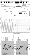
Figure 1. Organization and variation of DXZ4.
(A) Ideogram of the human X chromosome showing the location of DXZ4 at Xq23. Beneath this is a schematic representation of the region immediately surrounding DXZ4. The macrosatellite array and distal inverted monomers are represented by the arrow heads. The nearest gene PLS3 is indicated proximal to the array. (B) Representation of a single 3.0 kb DXZ4 monomer defined by HindIII. The internal microsatellite repeats are indicated as is the DXZ4 promoter region. (C) Predicted higher-order organization of the array as revealed by dot-plot analysis. A single 3.0 kb monomer sequence is on the y-axis, whereas the 120 kb genomic interval containing the array and inverted monomers is on the x-axis. The 120 kb sequence is located at 114.9 Mb on the human X chromosome (coordinates according to build hg19). Dot-plot generated using NCBI Blast, and the output image labeled in Adobe Photoshop CS2. (D) Copy number variation of DXZ4. Southern blot analysis of XbaI cut DNA from 22 unrelated individuals separated by pulsed field gel electrophoresis and hybridized with a DXZ4 probe. The ethnicity of the individuals used is indicated at the top. Size in kb is given to the right of each blot. The numbers given to the left of the blots with the double-headed arrow indicates the range of inferred DXZ4 copy number, with 12 in the smallest array and approximately 96 in the largest array.
Figure 2
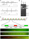
Figure 2. Confirmation of tandem arrangement for DXZ4.
(A) Predicted restriction endonuclease map for DXZ4 BAC clone 2272M5 using BamHI, B top, or HindIII, H bottom. Predicted fragment sizes given are in kb. The grey right facing arrows represent single 3 kb DXZ4 monomers. The central bracketed monomer represents all other tandem arranged monomers in the BAC. The large looped arrow (11.0 or 9.3) represents the pBeloBAC11 vector backbone. (B) Ethidium bromide stained agarose gel showing restriction fragments obtained from BAC 2272M5 when digested with either BamHI or HindIII. Fragment sizes are given to the right. (C) Tandem arrangement of DXZ4 in vivo as determined by extended fiber FISH. At the top is a predicted schematic for DXZ4 tandem arrangement, with each right facing arrow representing a 3.0 kb DXZ4 monomer. The alternating red and green circles represent probe locations. Beneath this is a representation of a single DXZ4 monomer indicating the location of the two probes used for fiber FISH of 550 bp (Green) and 449 bp (Red) separated by 899 bp or 1098 bp for the adjacent monomer. At the bottom are examples of merged Red and Green fluorescent images of extended DNA fiber hybridizations. Yellow signals indicate overlapping red and green probes in regions where fibers are not stretched to the same extent as the rest of the fiber.
Figure 3
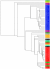
Figure 3. Relationship of DXZ4 monomer sequence within and between individuals.
Cladogram of 60 complete DXZ4 monomer DNA sequences. Tree image generated using MUSCLE version 3.8 . Color highlights added in Adobe Photoshop CS (Ver.8.0). Sequences labeled DXZ4-1 through 88 and highlighted in red represent sequences derived from BAC clone 2272M5 (this manuscript). Sequences labeled WI2 and highlighted in blue represent sequences taken from clones from the WIBR-2 human fosmid library. Sequence accession numbers include WI2-7 AC196704.1, WI2 AC193162 and WI2-8 CR753863.9. Sequences labeled ABC and colored green, mauve, orange and black are derived from fosmid libraries from four individuals of different ethnicities . Sequence accession numbers include: ABC9 AC212298.1, ABC11 AC236928.2, ABC13 AC226798 and ABC16 AC238719.3. The sequence highlighted in yellow annotated Giacalone is taken from accession number S60754 . Sequences labeled XXyac highlighted in grey are derived from XXyac-74A3 (BX546444.14). RP13 sequences derived from accession number AL392170.7.
Figure 4
Figure 4. DXZ4 inheritance and stability.
Inheritance of DXZ4 through three generations in three independent CEPH Utah pedigrees. (A) CEPH-1331, (B) CEPH-1333 and (C) CEPH-1345. Members of each family are indicated above the blot in the pedigrees and the members are given the Coriell GM0- ID for each member of the family. Hybridizing fragments are given to the left and right sides of the blots. The asterisks indicate alleles of altered size.
Figure 5
Figure 5. Expression of DXZ4.
(A) Examples showing the distribution of DXZ4 RNA versus XIST RNA in various 46,XX cells and a 46,XY cell line. The cell identity is indicated above each panel of three images. XIST RNA is shown in red, DXZ4 in green and nuclei are stained with DAPI (blue) in the merged image. (B) Expression of DXZ4 from the Xa and Xi in ten independent female lymphoblast cell lines given on the x-axis. Number of nuclei are given on the y-axis as percent. One hundred nuclei were scored for each cell line. (C) Detection of distinct regions of DXZ4 RNA by RNA FISH. The right facing arrow represents a single DXZ4 monomer with the regions highlighted in black (1–4) indicating the location of the probes used. Beneath this are examples of RNA FISH signals for each probe merged with DAPI. (D) RT-PCR analysis of DXZ4 using cDNA generated from total RNA isolated from 20 different human tissues. The tissues are listed above each agarose gel image, with a “+” indicating cDNA with reverse transcriptase and “−” indicating the no reverse transcriptase control. The top row indicates DXZ4 (labeled “D”) whereas the lower lane indicates GAPDH (labeled “G”). BM – bone marrow; Ce – cerebellum; WB – whole brain; FB – fetal brain; FL – fetal liver; H – heart; Li – liver; Lu – lung; P – prostate; SG – salivary gland; SM – skeletal muscle; Sp – spleen; Te – testis; Th – thymus; Tr – trachea; U – uterus; Co- colon; SI – small intesitine; SC – spinal cord; St – stomach.
Figure 6
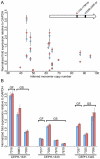
Figure 6. Lack of correlation between array size, expression and inheritance.
(A) Graph showing normalized expression levels of DXZ4 relative to GAPDH (y-axis) plotted against the inferred monomer copy number of DXZ4 (x-axis) for 22 different males as determined by PFGE. The right facing arrow represents a single DXZ4 monomer with the location of the regions amplified indicated. The plotted data indicates qRT-PCR for each of the two regions (pink diamond v blue diamond) from triplicate amplifications. (B) Expression levels of DXZ4 in maternal grandfathers (GF) plotted alongside grandsons (GS) who inherited their maternal grandfathers DXZ4 array. Data shown for three independent CEPH families.

References

    1. Lander ES, Linton LM, Birren B, Nusbaum C, Zody MC, et al. Initial sequencing and analysis of the human genome. Nature. 2001;409:860–921. - PubMed
    1. Warburton PE, Hasson D, Guillem F, Lescale C, Jin X, et al. Analysis of the largest tandemly repeated DNA families in the human genome. BMC Genomics. 2008;9:533. - PMC - PubMed
    1. Bruce HA, Sachs N, Rudnicki DD, Lin SG, Willour VL, et al. Long tandem repeats as a form of genomic copy number variation: structure and length polymorphism of a chromosome 5p repeat in control and schizophrenia populations. Psychiatr Genet. 2009;19:64–71. - PMC - PubMed
    1. Chadwick BP. DXZ4 chromatin adopts an opposing conformation to that of the surrounding chromosome and acquires a novel inactive X-specific role involving CTCF and antisense transcripts. Genome Res. 2008;18:1259–1269. - PMC - PubMed
    1. Giacalone J, Friedes J, Francke U. A novel GC-rich human macrosatellite VNTR in Xq24 is differentially methylated on active and inactive X chromosomes. Nat Genet. 1992;1:137–143. - PubMed

Publication types