Optical assessment of tumor resection margins in the breast
- PMID: 21544237
- PMCID: PMC3085495
- DOI: 10.1109/jstqe.2009.2033257
Optical assessment of tumor resection margins in the breast
Abstract
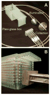
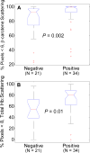
Breast conserving surgery, in which the breast tumor and surrounding normal tissue are removed, is the primary mode of treatment for invasive and in situ carcinomas of the breast, conditions that affect nearly 200,000 women annually. Of these nearly 200,000 patients who undergo this surgical procedure, between 20-70% of them may undergo additional surgeries to remove tumor that was left behind in the first surgery, due to the lack of intra-operative tools which can detect whether the boundaries of the excised specimens are free from residual cancer. Optical techniques have many attractive attributes which may make them useful tools for intra-operative assessment of breast tumor resection margins. In this manuscript, we discuss clinical design criteria for intra-operative breast tumor margin assessment, and review optical techniques appied to this problem. In addition, we report on the development and clinical testing of quantitative diffuse reflectance imaging (Q-DRI) as a potential solution to this clinical need. Q-DRI is a spectral imaging tool which has been applied to 56 resection margins in 48 patients at Duke University Medical Center. Clear sources of contrast between cancerous and cancer-free resection margins were identified with the device, and resulted in an overall accuracy of 75% in detecting positive margins.
Figures


References
-
- SUROS News Release. New Method for Breast Cancer Diagnosis. 2003.
-
- Tartter PI, Kaplan J, Bleiweiss I, Gajdos C, Kong A, Ahmed S, Zapetti D. Lumpectomy margins, reexcision, and local recurrence of breast cancer. Am J Surg. 2000 Feb;179:81–5. - PubMed
-
- Kunos C, Latson L, Overmoyer B, Silverman P, Shenk R, Kinsella T, Lyons J. Breast conservation surgery achieving >or=2 mm tumor-free margins results in decreased local-regional recurrence rates. Breast J. 2006 Jan-Feb;12:28–36. - PubMed
-
- Elkhuizen PH, van de Vijver MJ, Hermans J, Zonderland HM, van de Velde CJ, Leer JW. Local recurrence after breast-conserving therapy for invasive breast cancer: high incidence in young patients and association with poor survival. Int J Radiat Oncol Biol Phys. 1998 Mar 1;40:859–67. - PubMed
-
- Balch GC, Mithani SK, Simpson JF, Kelley MC. Accuracy of intraoperative gross examination of surgical margin status in women undergoing partial mastectomy for breast malignancy. Am Surg. 2005 Jan;71:22–7. discussion 27–8. - PubMed
Grants and funding
LinkOut - more resources
Full Text Sources
Other Literature Sources
