

Biofilm formation and adherence characteristics of an Elizabethkingia meningoseptica isolate from Oreochromis mossambicus
- PMID: 21545730
- PMCID: PMC3112384
- DOI: 10.1186/1476-0711-10-16
Biofilm formation and adherence characteristics of an Elizabethkingia meningoseptica isolate from Oreochromis mossambicus
Abstract
Background: Elizabethkingia spp. are opportunistic pathogens often found associated with intravascular device-related bacteraemias and ventilator-associated pneumonia. Their ability to exist as biofilm structures has been alluded to but not extensively investigated.
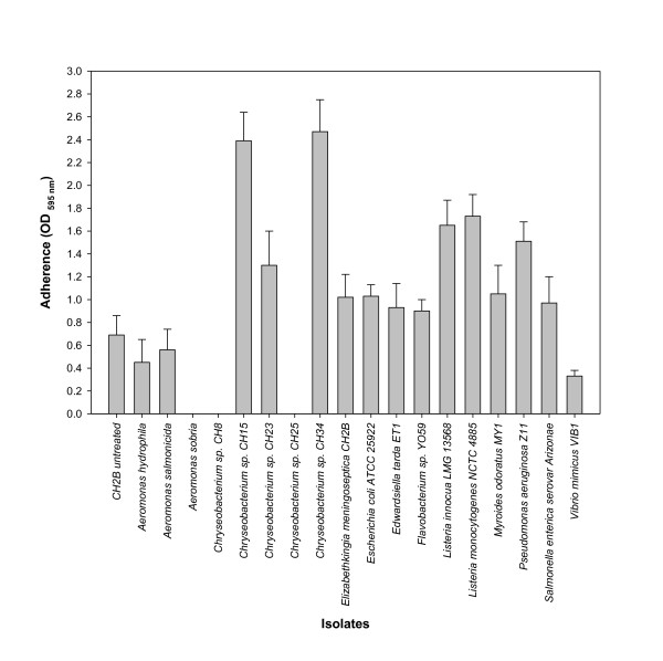
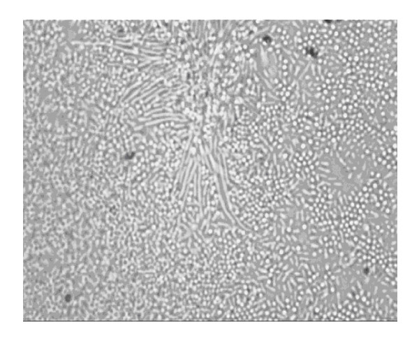
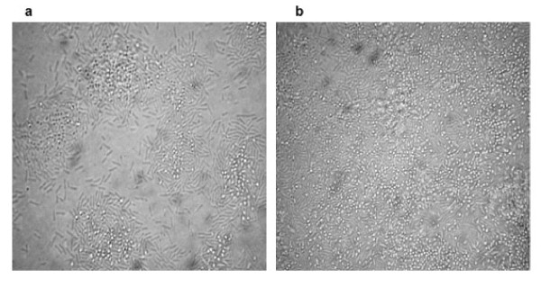
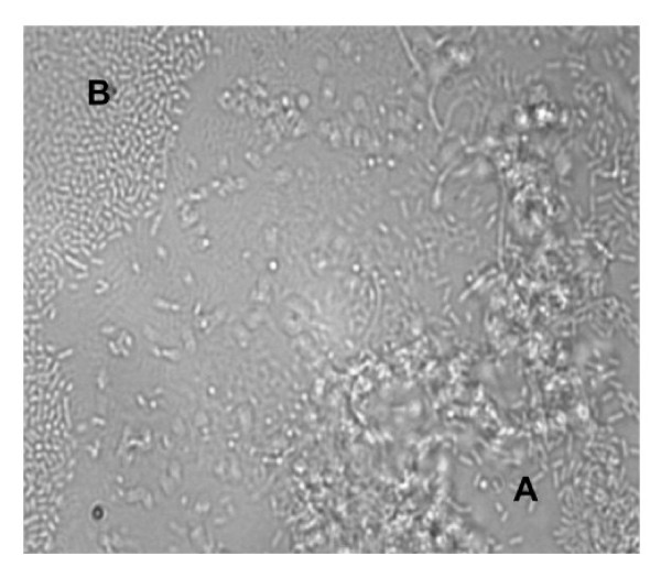
Methods: The ability of Elizabethkingia meningoseptica isolate CH2B from freshwater tilapia (Oreochromis mossambicus) and E. meningoseptica strain NCTC 10016(T) to adhere to abiotic surfaces was investigated using microtiter plate adherence assays following exposure to varying physico-chemical challenges. The role of cell-surface properties was investigated using hydrophobicity (bacterial adherence to hydrocarbons), autoaggregation and coaggregation assays. The role of extracellular components in adherence was determined using reversal or inhibition of coaggregation assays in conjunction with Listeria spp. isolates, while the role of cell-free supernatants, from diverse bacteria, in inducing enhanced adherence was investigated using microtitre plate assays. Biofilm architecture of isolate CH2B alone as well as in co-culture with Listeria monocytogenes was investigated using flow cells and microscopy.
Results: E. meningoseptica isolates CH2B and NCTC 10016(T) demonstrated stronger biofilm formation in nutrient-rich medium compared to nutrient-poor medium at both 21 and 37°C, respectively. Both isolates displayed a hydrophilic cell surface following the bacterial adherence to xylene assay. Varying autoaggregation and coaggregation indices were observed for the E. meningoseptica isolates. Coaggregation by isolate CH2B appeared to be strongest with foodborne pathogens like Enterococcus, Staphylococcus and Listeria spp. Partial inhibition of coaggregation was observed when isolate CH2B was treated with heat or protease exposure, suggesting the presence of heat-sensitive adhesins, although sugar treatment resulted in increased coaggregation and may be associated with a lactose-associated lectin or capsule-mediated attachment.
Conclusions: E. meningoseptica isolate CH2B and strain NCTC 10016(T) displayed a strong biofilm-forming phenotype which may play a role in its potential pathogenicity in both clinical and aquaculture environments. The ability of E. meningoseptica isolates to adhere to abiotic surfaces and form biofilm structures may result from the hydrophilic cell surface and multiple adhesins located around the cell.
Figures
References
-
- Bernardet J-F, Hugo C, Bruun B. In: The Prokaryotes. 3. Dworkin M, Falkow S, Rosenberg E, Schleifer K-H, Stackebrandt E, editor. Vol. 10. New York: Springer; 2006. The genera Chryseobacterium and Elizabethkingia; pp. 638–676.
-
- Lee C-H, Lin W-C, Chia J-H, Su L-H, Chien C-C, Mao A-H, Liu J-W. Community-acquired osteomyelitis caused by Chryseobacterium meningosepticum: case report and literature review. Diagn Microbiol Infect Dis. 2008;10:89–93. - PubMed
-
- Michel C, Matte-Tailliez O, Kerouault B, Bernardet J-F. Resistance pattern and assessment of phenicol agents' minimum inhibitory concentration in multiple drug resistant Chryseobacterium isolates from fish and aquatic habitats. J Appl Microbiol. 2005;10:323–332. doi: 10.1111/j.1365-2672.2005.02592.x. - DOI - PubMed
-
- Lin P-Y, Chen H-L, Huang C-T, Su L-H, Chiu C-H. Biofilm production, use of indwelling catheters and inappropriate antimicrobial therapy as predictors of fatality in Chryseobacterium meningosepticum bacteraemia. Int J Antimicrob Agent. 2010;10:436–440. doi: 10.1016/j.ijantimicag.2010.06.033. - DOI - PubMed
Publication types
MeSH terms
LinkOut - more resources
Full Text Sources
Molecular Biology Databases
Miscellaneous
