. 2011 Jul 1;355(1):152-62.
doi: 10.1016/j.ydbio.2011.04.025. Epub 2011 Apr 28.

Hedgehog signaling controls homeostasis of adult intestinal smooth muscle

Affiliations

Hedgehog signaling controls homeostasis of adult intestinal smooth muscle

William J Zacharias et al. Dev Biol. .

Abstract

The Hedgehog (Hh) pathway plays multiple patterning roles during development of the mammalian gastrointestinal tract, but its role in adult gut function has not been extensively examined. Here we show that chronic reduction in the combined epithelial Indian (Ihh) and Sonic (Shh) hedgehog signal leads to mislocalization of intestinal subepithelial myofibroblasts, loss of smooth muscle in villus cores and muscularis mucosa as well as crypt hyperplasia. In contrast, chronic over-expression of Ihh in the intestinal epithelium leads to progressive expansion of villus smooth muscle, but does not result in reduced epithelial proliferation. Together, these mouse models show that smooth muscle populations in the adult intestinal lamina propria are highly sensitive to the level of Hh ligand. We demonstrate further that Hh ligand drives smooth muscle differentiation in primary intestinal mesenchyme cultures and that cell-autonomous Hh signal transduction in C3H10T1/2 cells activates the smooth muscle master regulator Myocardin (Myocd) and induces smooth muscle differentiation. The rapid kinetics of Myocd activation by Hh ligands as well as the presence of an unusual concentration of Gli sties in this gene suggest that regulation of Myocd by Hh might be direct. Thus, these data indicate that Hh is a critical regulator of adult intestinal smooth muscle homeostasis and suggest an important link between Hh signaling and Myocd activation. Moreover, the data support the idea that lowered Hh signals promote crypt expansion and increased epithelial cell proliferation, but indicate that chronically increased Hh ligand levels do not dampen crypt proliferation as previously proposed.


Figures

Figure 1
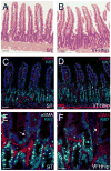
Figure 1. Crypt hyperplasia and mislocalized myofibroblasts in VHhip mice
A,B) Histological (H&E) analysis of 3 month old single transgenic (ST, A) and VFHhip (DTG, B) animals. Note increased crypt depth. C–F) Immunostaining for αSMA (red, myofibroblast and SMC marker) and Ki67 (green, proliferation marker) in VFHhip mice and ST littermates. Proliferative epithelial cells fill the elongated crypts and are located on villus tips (D,F) in VFHhip animals. Subepithelial myofibroblasts are found farther up the crypt/villus axis in response to Hh inhibition in 1VFHhip animals, arrows, (F). Bars: A–D=50μm; E–F=20μm.
Figure 2
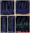
Figure 2. Loss of differentiated SMC in VHhip mice
A–D) Immunostaining for αSMA (red) and desmin (green) reveals differentiated SMC (yellow) in the cores of villi of ST animals (A,C), but not in VFHhip animals at 3 months. SMC precursors (desmin positive only, green) are expanded in the cores of VFHhip mice (B,D). E) Focal loss of muscularis mucosa in VFHhip animals (arrows). This section is stained with αSMA and Ki67 (green, proliferation marker). Bars: A–C=50μm; C–E=20μm
Figure 3
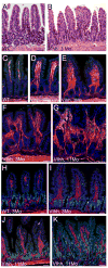
Figure 3. Progressive expansion of villus SMC and distortion of villus and crypt architecture in VIhh mice
A,B) Histological (H&E) analysis of 3 month old WT (A) animals and VIhh (B) littermates. C–G) αSMA (red) and desmin (green) staining in WT mice (C) and VIhh mice (D–G) at ages marked on panels. Though all VIhh villi show expansion of SMC cores, areas of major disortion are focal (F,G) and increase with age. H–K) Sections are stained with αSMA (red) and Ki67 (green). At 3 months of age there is no evidence of difference in epithelial proliferation between WT mice (H) and VIhh mice (I). Later in life, the majority of the epithelium have unaltered proliferation (J), but in areas of major distortion of crypt/villus architecture, robust epithelial proliferation is seen (K). Bars=50μm.
Figure 4
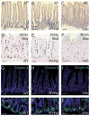
Figure 4. Transgenic modulation of Hh signaling does not alter epithelial lineage allocation at 3 months of age
A–C) Alkaline Phosphatase (AP) staining to visulaize the absorptive surface of enterocytes. The absorptive surface is reduced by expanded crypts in VFHhip animals (B) but is otherwise normal. VIhh animals (C) have similar absorptive surface to WT (A). D–F) PAS/Alcian Blue stain to visualize goblet cells. Both VFHhip (E) and VIhh (F) animals have a similar number of goblet cells compared to WT (D). G–H) Chromogranin A (Chromo A) stain for endocrine cells, with DAPI nuclear conterstain. Neither inhibition of Hh (H) or overexpression of Hh (I) alter enteroendocrine cell number or position. J–L) Lysozyme (Lyso) staining for detection of Paneth cells, showing high magnification of crypts. No change in Paneth cell number or location is seen with Hh modulation. A–I shown at 200× magnification, J–K at 600×.
Figure 5
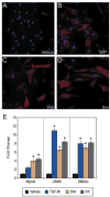
Figure 5. Hh treatment of E18.5 mesenchyme causes Hh pathway activation, increases in myofibroblasts and SMC differentiation
A–C) Activation of Hh signaling in E18.5 Gli1+/lacZ mesenchyme. (A) Mesenchyme was cultured for 48 hours and treated with vehicle for another 24 hours. Lack of β-galactosidase activity reflects lack of active Hh signal transduction. B,C) Mesenchymal samples were held in culture for 48 hours, then treated for 24 hours with Shh (B) or Ihh (C). Hh signal transduction is activated. D) αSMA (red) and desmin (green) immunofluorescence demonstrates that myofibroblasts (red), SMC precursor cells (green) and differentiated SMC (yellow) are present in untreated isolated mesenchyme. E) Quantitation of myofibroblasts and SMC populations after Hh treatment in isolated mesenchyme. Shown are SMC precursors (Desmin), myofibroblasts (αSMA), and SMC (αSMA/Desmin) as a percentage of all cells after 24 hour exposure to vehicle, Shh or Ihh. Treatment with Hh ligand decreases precursor cells and increases myofibroblasts and differentiated SMC cells. * = p<0.05 by Student's t-test compared to vehicle treatment.
Figure 6
Figure 6. Hh ligands induce Myocd with kinetics similar to known Hh target genes.D
A) Q-RT-RCR of Bmp4 and Myocd expression in VIhh (left panel) and VFHhip animals (right panel). * = p<0.05 compared to WT or ST mice. Reduction in Myocd level of VFHhip animals is not significant, likely because ME is not affected in this model. B) Q-RT-PCR of Myocd and several Hh target genes after exposure of isolated intestinal mesenchyme to Shh or Ihh for 24 hours. (Mesenchyme was held in culture for 48 hours prior to treatment with Hh ligands). C,D) Time course of target gene expression after Shh (C) or Ihh (D) treatment. The kinetics of Myocd activation (purple line) most closely parallels the direct Hh target, Bmp4 (green line). Igf-1 (yellow line), also expressed by SMC, is not activated after 24 hours of treatment with Hh ligand.
Figure 7
Figure 7. Hh promotes SMC differentiation in C3H10T1/2 cells in the presence of low serum
A) 10T1/2 cells do not express αSMA when treated with vehicle in the presence of low serum. B) Tgfβ1 induces αSMA expression and morphological change in 10T1/2 cells. C,D) Shh (C) and Ihh (D) treatment drive SMC differentiation as indicated by αSMA expression and cell shape change in a manner similar to Tgfβ1. G) Q-RT-PCR analysis of SMC markers in 10T1/2 cells after treatment with Tgfβ1 or Hh ligand. * = p < 0.05 by Student's t-test.
Figure 8
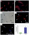
Figure 8. Hh pathway activation is sufficient to induce cell autonomous SMC differentiation of C3H10T1/2 cells
A–B) Treatment of cells with Shh (A) or Ihh (B) in the presence of the Bmp-inhibitor, Noggin, does not alter Hh-induced αSMA expression in 10T1/2 cells (compare to Figure 7C–D), indicating that Hh does not require BMP signaling to induce SMC differentiation. C–E) Gli2ΔN-transfected 10T1/2 cells exposed to differentiation media. C) DIC image; five cells are marked with arrows. D) Two cells express αSMA and take on an SMC-like morphology, whereas adjacent untransfected cells do not express αSMA. E) Staining for the Myc epitope tag confirms that these cells are positive for Gli2ΔN. F) Quantification of smooth muscle differentiation after transfection with GFP or Gli2ΔN. Ten fields from four wells in two transfection experiments were counted. * = p<0.01 by Student’s t-test.

References

    1. Antonucci A, Fronzoni L, Cogliandro L, Cogliandro RF, Caputo C, De Giorgio R, Pallotti F, Barbara G, Corinaldesi R, Stanghellini V. Chronic intestinal pseudo-obstruction. World J Gastroenterol. 2008;14:2953–61. - PMC - PubMed
    1. Bai CB, Auerbach W, Lee JS, Stephen D, Joyner AL. Gli2, but not Gli1, is required for initial Shh signaling and ectopic activation of the Shh pathway. Development. 2002;129:4753–61. - PubMed
    1. Caubit X, Lye CM, Martin E, Core N, Long DA, Vola C, Jenkins D, Garratt AN, Skaer H, Woolf AS, et al. Teashirt 3 is necessary for ureteral smooth muscle differentiation downstream of SHH and BMP4. Development. 2008;135:3301–10. - PubMed
    1. Chen J, Chen H, Sanders KM, Perrino BA. Regulation of SRF/CArG-dependent gene transcription during chronic partial obstruction of murine small intestine. Neurogastroenterol Motil. 2008;20:829–42. - PMC - PubMed
    1. Choudhury BK, Shi XZ, Sarna SK. Gene plasticity in colonic circular smooth muscle cells underlies motility dysfunction in a model of postinfective IBS. Am J Physiol Gastrointest Liver Physiol. 2009;296:G632–42. - PMC - PubMed

Publication types

MeSH terms