. 2011 Jul 1;51(1):216-24.
doi: 10.1016/j.freeradbiomed.2011.04.026. Epub 2011 Apr 19.

Nrf2 has a protective role against neuronal and capillary degeneration in retinal ischemia-reperfusion injury

Affiliations

Nrf2 has a protective role against neuronal and capillary degeneration in retinal ischemia-reperfusion injury

Yanhong Wei et al. Free Radic Biol Med. .

Abstract

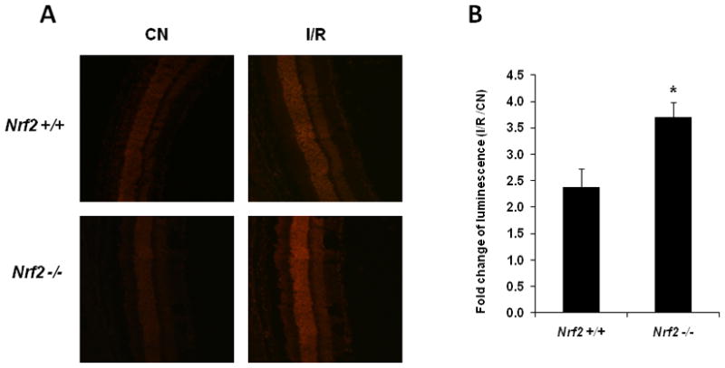
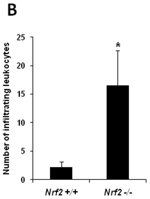
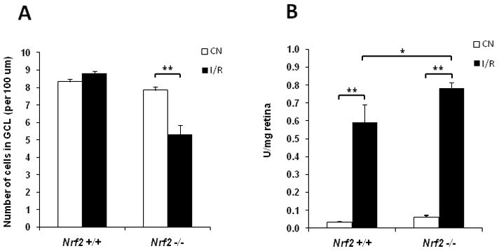
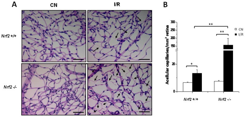
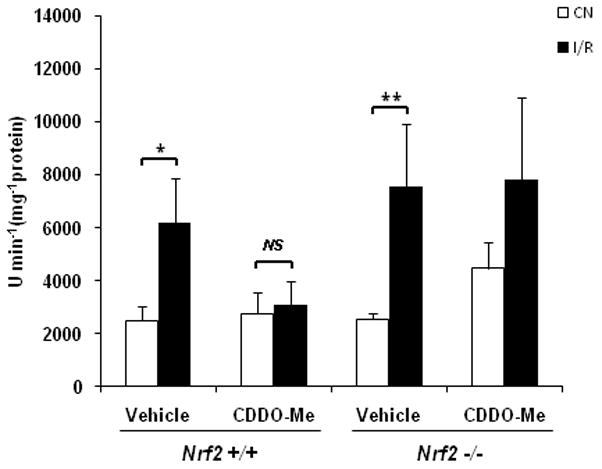
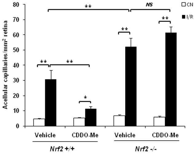
Retinal ischemia-reperfusion (I/R) involves an extensive increase in reactive oxygen species as well as proinflammatory changes that result in significant histopathologic damage, including neuronal and vascular degeneration. Nrf2 has a well-known cytoprotective role in many tissues, but its protective function in the retina is unclear. We investigated the possible role of Nrf2 as a protective mechanism in retinal ischemia-reperfusion injury using Nrf2(-/-) mice. I/R resulted in an increase in retinal levels of superoxide and proinflammatory mediators, as well as leukocyte infiltration of the retina and vitreous, in Nrf2(+/+) mice. These effects were greatly accentuated in Nrf2(-/-) mice. With regard to histopathologic damage, Nrf2(-/-) mice exhibited loss of cells in the ganglion cell layer and markedly accentuated retinal capillary degeneration, as compared to wild-type. Treatment with the Nrf2 activator CDDO-Me increased antioxidant gene expression and normalized I/R-induced superoxide in the retina in wild-type but not Nrf2(-/-) mice. CDDO-Me treatment abrogated retinal capillary degeneration induced by I/R in wild-type but not Nrf2(-/-) mice. These studies indicate that Nrf2 is an important cytoprotective mechanism in the retina in response to ischemia-reperfusion injury and suggest that pharmacologic induction of Nrf2 could be a new therapeutic strategy for retinal ischemia-reperfusion and other retinal diseases.


Figures

Fig. 1
Fig. 1. Nrf2-deficient mice exhibit accentuated induction of superoxide levels after retinal I/R injury
Retinas were obtained and analyzed 24 hrs after I/R. (A) DHE fluorescence was significantly increased in Nrf2 +/+ retinas, but were even greater in retinas from Nrf2 −/− mice following I/R injury. Representative images from each group are shown (n = 4 – 5). (B) Retinal superoxide levels were determined by biochemical assay using lucigenin. Data are expressed as fold-change of luminescence in the retina subjected to I/R vs. control retina (contralateral eye, not subjected to I/R) (n = 4 – 5). The increase in luminescence induced by I/R was significantly greater in Nrf2 −/− mice. CN, control retinas without I/R. * p < 0.05.
Fig. 2
Fig. 2. Deletion of Nrf2 increases mRNA expression of inflammatory mediators in the retina after I/R injury
Real-time PCR analysis of inflammatory mediators was performed on retinas that were harvested 6 hours after I/R. Data are expressed as fold-change in the retina subjected to I/R vs. control (CN) retina (contralateral eye, not subjected to I/R). n = 5, * p < 0.05; ** p < 0.01.
Fig. 3
Fig. 3. Deletion of Nrf2 promotes more extensive inflammation after retinal I/R injury
Eyes were harvested and subjected to H&E staining for histological analysis 2 days after I/R. (A) The histological features of the retina from control (non-I/R) wild-type and Nrf2 −/− mice were similar and showed no significant histological changes (a and d). Wild-type mice exhibited leukocyte (predominantly neutrophil) infiltration in the vitreous (short arrow) after I/R injury (b and c). The severity of inflammation was far more extensive after I/R in Nrf2 −/− mice (e and f), with significantly greater numbers of neutrophils (short arrow and inset of panel f). There was detachment of the internal limiting membrane (ILM) (long arrow) after I/R in all the Nrf2 −/− mice, and many neutrophils accumulated in this space (short arrow). Fig. 3c and 3f are higher magnification images of the enclosed area in Fig. 3b and 3e, respectively. Scale bar, 50 μm. (B) Infiltrating leukocytes were quantitated in 10 random high-powered fields for each eye, as described in Methods. The number of infiltrating leukocytes was significantly higher in Nrf2 −/− than wild-type retina after I/R injury. n = 5, * p < 0.05.
Fig. 4
Fig. 4. Nrf2-deficient mice exhibit more pronounced neuronal cell loss after retinal I/R injury
(A) Ganglion cell layer counting was performed as described in Methods. The number of cells in ganglion cell layer (GCL) of the I/R retina was significantly less than that of control retina (CN) in Nrf2 −/−mice 2 days after I/R injury, while no significant change was observed in the wild-type mice (n = 5, ** p < 0.01). (B) Apoptotic DNA Cleavage ELISA was performed 48 hrs after I/R injury. Nucleosomal DNA fragmentation was significantly increased in wild-type mice after I/R injury. There was a further increase in apoptotic DNA cleavage in Nrf2 −/− mice after I/R injury, beyond what was observed in wild-type mice. n = 5, * p < 0.05; ** p < 0.01.
Fig. 5
Fig. 5. Retinal capillary degeneration following I/R injury is markedly accentuated in Nrf2-deficient mice
Retinas were subjected to trypsin digest to visualize the microvasculature and quantify acellular capillaries 8 days after I/R. (A) Following I/R injury, Nrf2 +/+ retinas exhibited increased number of acellular capillaries (arrows). Nrf2 −/− retinas exhibited a greatly accentuated increase in acellular capillaries following I/R injury. Scale bar, 100 μm. (B) Quantification of acellular capillaries normalized by the counting area. CN, control retinas without I/R. n = 4 – 5, * p < 0.05; ** p < 0.01.
Fig. 6
Fig. 6. CDDO-Me increases the expression of Nrf2-responsive antioxidant genes in wild-type, but not Nrf2 −/− retinas
Mice were treated with one intraperitoneal injection of 1 μmol/kg CDDO-Me or vehicle 6 hrs before isolating the retina. n = 4, * p < 0.05; NS: not significant.
Fig. 7
Fig. 7. CDDO-Me suppresses superoxide increase in wild-type but not Nrf2 −/− retinas following I/R injury
Mice were pre-treated with three intraperitoneal injections of 1 μmol/kg CDDO-Me or vehicle at 48, 24 and 0 hr before being subjected to I/R. Retinal superoxide levels were determined by biochemical assay using lucigenin 24 hrs after I/R. CDDO-Me treatment completely abrogated the increase in retinal superoxide levels induced by I/R in wild-type mice. CDDO-Me treatment had no effect on this I/R-induced increase in retinal superoxide in Nrf2 −/− mice. CN, control retinas without I/R. n = 5, * p < 0.05; ** p < 0.01; NS: not significant.
Fig. 8
Fig. 8. CDDO-Me inhibits retinal capillary degeneration in wild-type but not Nrf2 −/− retinas following I/R injury
Mice were pre-treated with three intraperitoneal injections of 1 μmol/kg CDDO-Me or vehicle at 48, 24 and 0 hr before being subjected to I/R. After I/R, the mice were treated with 1 μmol/kg CDDO-Me or vehicle every 48 hours. 8 days after I/R, retinas were subjected to trypsin digest to visualize the microvasculature and quantify acellular capillaries. Treatment with CDDO-Me markedly reduced the number of acellular capillaries in wild-type retina, but showed no beneficial effect on Nrf2 −/− retinas. CN, control retinas without I/R. n = 4 – 5, * p < 0.05; ** p < 0.01; NS: not significant.

References

    1. Osborne NN, Casson RJ, Wood JP, Chidlow G, Graham M, Melena J. Retinal ischemia: mechanisms of damage and potential therapeutic strategies. Prog Retin Eye Res. 2004;23:91–147. - PubMed
    1. McCord JM. Oxygen-derived free radicals in postischemic tissue injury. N Engl J Med. 1985;312:159–163. - PubMed
    1. Zweier JL, Flaherty JT, Weisfeldt ML. Direct measurement of free radical generation following reperfusion of ischemic myocardium. Proc Natl Acad Sci U S A. 1987;84:1404–1407. - PMC - PubMed
    1. Korthuis RJ, Granger DN. Reactive oxygen metabolites, neutrophils, and the pathogenesis of ischemic-tissue/reperfusion. Clin Cardiol. 1993;16:I19–26. - PubMed
    1. Hangai M, Yoshimura N, Hiroi K, Mandai M, Honda Y. Inducible nitric oxide synthase in retinal ischemia-reperfusion injury. Exp Eye Res. 1996;63:501–509. - PubMed

Publication types

MeSH terms

LinkOut - more resources