

Complement in the brain
- PMID: 21546088
- PMCID: PMC3142281
- DOI: 10.1016/j.molimm.2011.04.003
Complement in the brain
Abstract
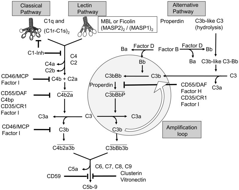
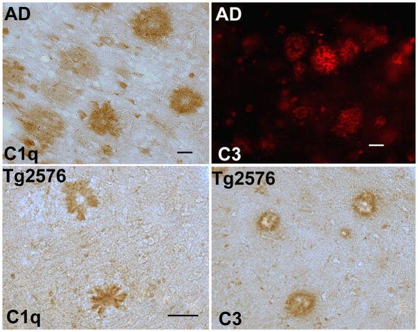
The brain is considered to be an immune privileged site, because the blood-brain barrier limits entry of blood borne cells and proteins into the central nervous system (CNS). As a result, the detection and clearance of invading microorganisms and senescent cells as well as surplus neurotransmitters, aged and glycated proteins, in order to maintain a healthy environment for neuronal and glial cells, is largely confined to the innate immune system. In recent years it has become clear that many factors of innate immunity are expressed throughout the brain. Neuronal and glial cells express Toll like receptors as well as complement receptors, and virtually all complement components can be locally produced in the brain, often in response to injury or developmental cues. However, as inflammatory reactions could interfere with proper functioning of the brain, tight and fine tuned regulatory mechanisms are warranted. In age related diseases, such as Alzheimer's disease (AD), accumulating amyloid proteins elicit complement activation and a local, chronic inflammatory response that leads to attraction and activation of glial cells that, under such activation conditions, can produce neurotoxic substances, including pro-inflammatory cytokines and oxygen radicals. This process may be exacerbated by a disturbed balance between complement activators and complement regulatory proteins such as occurs in AD, as the local synthesis of these proteins is differentially regulated by pro-inflammatory cytokines. Much knowledge about the role of complement in neurodegenerative diseases has been derived from animal studies with transgenic overexpressing or knockout mice for specific complement factors or receptors. These studies have provided insight into the potential therapeutic use of complement regulators and complement receptor antagonists in chronic neurodegenerative diseases as well as in acute conditions, such as stroke. Interestingly, recent animal studies have also indicated that complement activation products are involved in brain development and synapse formation. Not only are these findings important for the understanding of how brain development and neural network formation is organized, it may also give insights into the role of complement in processes of neurodegeneration and neuroprotection in the injured or aged and diseased adult central nervous system, and thus aid in identifying novel and specific targets for therapeutic intervention.
Copyright © 2011 Elsevier Ltd. All rights reserved.
Figures
References
-
- Abbott NJ, Patabendige AA, Dolman DE, Yusof SR, Begley DJ. Structure and function of the blood-brain barrier. Neurobiol Dis. 2010;37:13–25. - PubMed
-
- Afagh A, Cummings BJ, Cribbs DH, Cotman CW, Tenner AJ. Localization and cell association of C1q in Alzheimer's disease brain. Exp Neurol. 1996;138:22–32. - PubMed
-
- Akiyama H, McGeer PL. Brain microglia constitutively express B-2 integrins. J Neuroimmunol. 1990;30:81–93. - PubMed
-
- Austen KF, Fearon DT. A molecular basis of activation of the alternative pathway of human complement. Adv Exp Med Biol. 1979;120B:3–17. - PubMed
Publication types
MeSH terms
Substances
Grants and funding
LinkOut - more resources
Full Text Sources
Other Literature Sources
Research Materials
