. 2011 Jun;29(5):683-92.
doi: 10.1016/j.mri.2011.02.022. Epub 2011 May 5.

Accuracy of image registration between MRI and light microscopy in the ex vivo brain

Affiliations

Accuracy of image registration between MRI and light microscopy in the ex vivo brain

Ann S Choe et al. Magn Reson Imaging. 2011 Jun.

Abstract

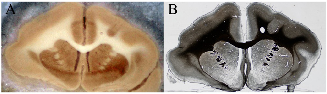
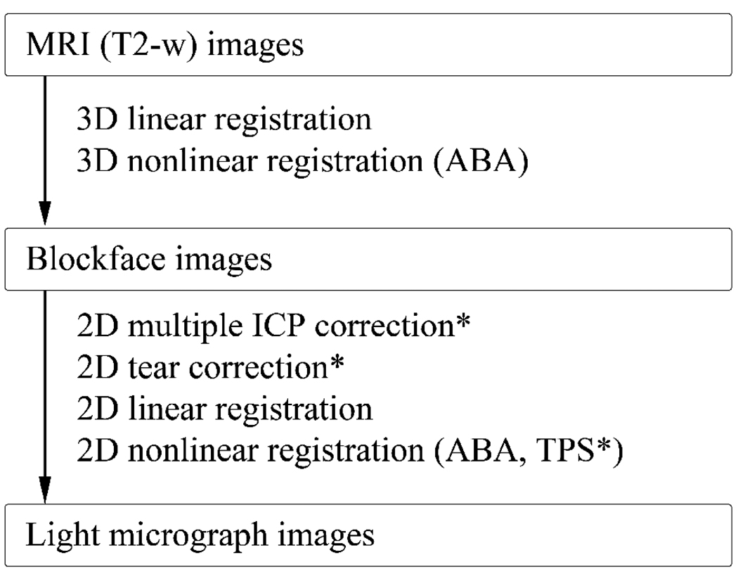
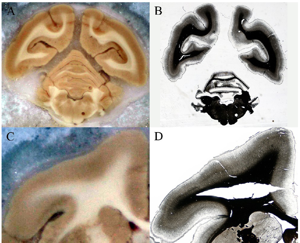
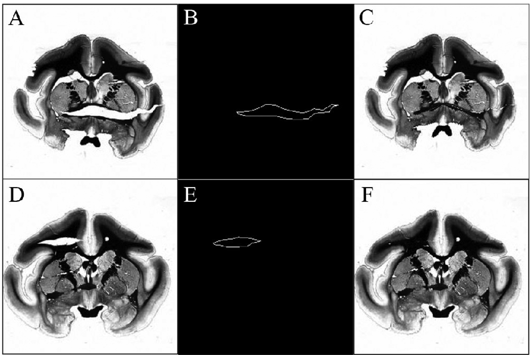
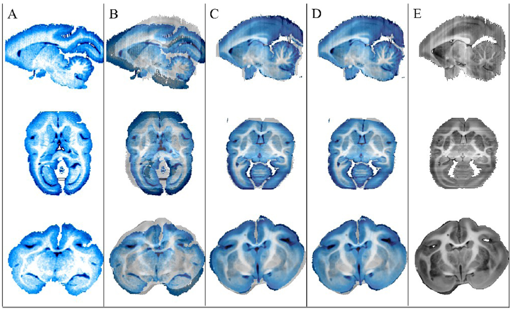
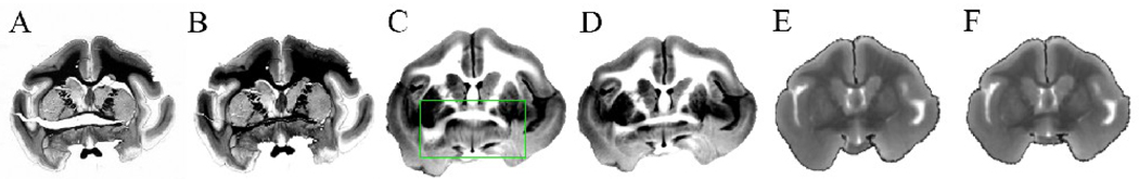
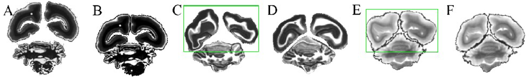
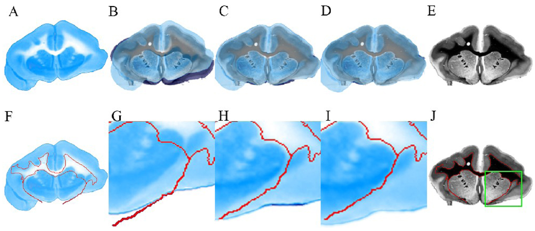
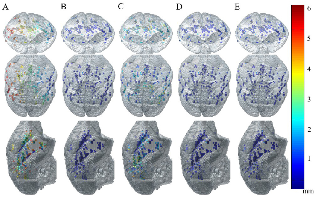
A multistep procedure was developed to register magnetic resonance imaging (MRI) and histological data from the same sample in the light microscopy image space, with the ultimate goal of allowing quantitative comparisons of the two datasets. The fixed brain of an owl monkey was used to develop and test the procedure. In addition to the MRI and histological data, photographic images of the brain tissue block acquired during sectioning were assembled into a blockface volume to provide an intermediate step for the overall registration process. The MR volume was first registered to the blockface volume using a combination of linear and nonlinear registration, and two dimensional (2D) blockface sections were registered to corresponding myelin-stained sections using a combination of linear and nonlinear registration. Before this 2D registration, two major types of tissue distortions were corrected: tissue tearing and independent movement of different parts of the brain, both introduced during histological processing of the sections. The correction procedure utilized a 2D method to close tissue tears and a multiple iterative closest point (ICP) algorithm to reposition separate pieces of tissue in the image. The accuracy of the overall MR to micrograph registration procedure was assessed by measuring the distance between registered landmarks chosen in the MR image space and the corresponding landmarks chosen in the micrograph space. The average error distance of the MR data registered to micrograph data was 0.324±0.277 mm, only 8% larger than the width of the MRI voxel (0.3 mm).


Figures

Fig 1
Fig 1
Blockface and histological images. (A) An example of a blockface image before the brain is segmented from its dry ice background (B) An example of a light micrograph image before the brain is segmented from its background. The section is the same as shown in (A), and is stained for myelin using the Gallyas silver staining method.
Fig 2
Fig 2
Multi-step registration workflow summary. Three major datasets – MRI, blockface, and light microscopy datasets – were acquired. The datasets were registered to each other using a combination of linear and nonlinear registration. For selected histological sections with tissue tearing or relative displacement of different parts of brain, 2D rigid tear correction and/or 2D multiple ICP correction was performed as a preprocessing step. In some cases, an additional step using TPS was necessary after nonlinear registration of the blockface images.
Fig 3
Fig 3
Image artifacts introduced during histological processing. (A), (C) Undistorted blockface images. (B) Example of relative displacement of the hemispheres and the cerebellum. (D) Example of tissue tearing.
Fig 4
Fig 4
Contour selection for 2Dtear correction. (A), (D) Original light micrographs with severe tissue tearing artifacts. (B), (E) User selected contours of tissue tear edges. (C), (F) Result of tear correction method.
Fig 5
Fig 5
3D registration of MRI (T2-w) to blockface volume. (A) Orthogonal views of the original non-diffusion weighted (T2-w) image volume. (B) Original T2-w images superimposed on blockface images, (C) T2-w images after linear registration, and (D) T2-w images after linear and nonlinear registration are overlaid on original blockface images, reproduced in (E).
Fig 6
Fig 6
Example of application of the tear correction method. (A) An original light micrograph, (B) the micrograph after closing the tear, (C) & (D) the corresponding blockface image deformed to match (A) and (B), respectively. (E) & (F) MR images registered to (A) & (B), respectively. Note the green region in (C) was locally stretched by the nonlinear registration algorithm in order to match the hole in (A), causing a distortion.
Fig 7
Fig 7
Example of application of the multiple component ICP method. (A) The original light micrograph of mounted tissue, (B) the corrected micrograph using the ICP algorithm, (C) & (D) the deformed blockface images and (E) & (F) MR images registered to (A) & (B), respectively. The green region shows the large distortion when the ICP algorithm was not applied to the light micrograph.
Fig 8
Fig 8
2D registration of a blockface section to a corresponding myelin stained section. (A) Original blockface section. (B) Original blockface section, (C) blockface section after linear registration, and (D) blockface section after linear and nonlinear registration overlaid on the myelin stained section, respectively. (E) Original myelin stained section. (F) Original blockface section. The red line represents the white matter (WM) outline of the corresponding myelin stained section, also outlined in (J). (G) A region of interest (ROI), outlined in green in (J), is selected from the original blockface section. (H) ROI in the blockface section after linear registration. (I) ROI in the blockface section after linear and nonlinear registration. In (G–I), the WM outline (in red) of the corresponding myelin stained section is also overlaid for comparison.
Fig 9
Fig 9
Distribution of landmarks used for registration accuracy measurements. Chosen landmarks are visualized within the surface rendered image of the original MRI volume. Each of the landmarks is color coded according to the registration error between landmarks in (A) original MRI image space and original blockface image space, (B) registered (linear & nonlinear) MRI image space and original blockface image space, (C) original blockface image space and original light micrograph space, (D) registered (linear & nonlinear) blockface image space and original light micrograph space, (E) registered (linear & nonlinear) MRI image space and original light micrograph space. The size of the voxels is 0.3 mm3.

References

    1. Blum F. Notiz uber die anwendung des formaldehyds (formol) als harrungs-und koservierungsmittel. Anat Anz. 1894;9:229.
    1. Underhill BML. The rate of penetration of fixatives. J R Microsc Soc. 1932;52:113.
    1. Durgun-Yucel B, Hopwood D, Yucel AH. The effects of mercaptoethanol-formaldehyde on tissue fixation and protein retention. Histochem J. 1996;28(5):375–383. - PubMed
    1. Fox CH, Johnson FB, Whiting J, Roller PP. Formaldehyde fixation. J Histochem Cytochem. 1985;33(8):845–853. - PubMed
    1. Bancroft JD, Cook HC. Manual of Histological Techniques and their Diagnositic Application. Edinburgh: New York Churchill Livingstone; 1994. pp. 349–356.

Publication types