. 2011 Oct;17(10):626-36.
doi: 10.1093/molehr/gar031. Epub 2011 May 5.

Prokineticin 1 induces Dickkopf 1 expression and regulates cell proliferation and decidualization in the human endometrium

Affiliations

Prokineticin 1 induces Dickkopf 1 expression and regulates cell proliferation and decidualization in the human endometrium

Linsay J Macdonald et al. Mol Hum Reprod. 2011 Oct.

Abstract

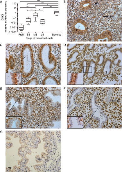
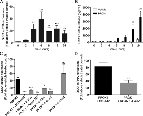
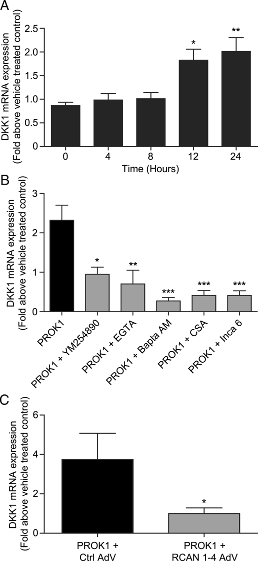
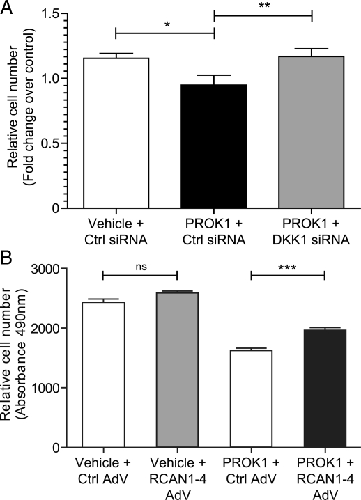
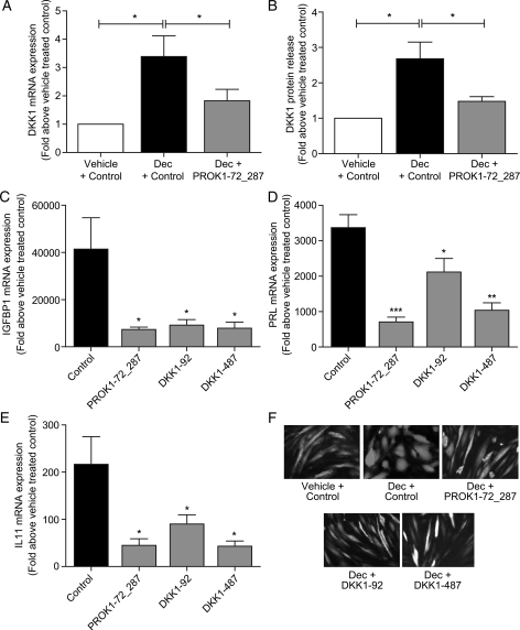
Prokineticin 1 (PROK1) signalling via prokineticin receptor 1 (PROKR1) regulates the expression of several genes with important roles in endometrial receptivity and implantation. This study investigated PROK1 regulation of Dickkopf 1 (DKK1) expression, a negative regulator of canonical Wnt signalling, and its function in the non-pregnant endometrium and first trimester decidua. DKK1 mRNA expression is elevated during the mid-secretory phase of the menstrual cycle and expression increases further in first trimester decidua. DKK1 protein expression is localized to glandular epithelial and stromal cells during the proliferative, early- and mid-secretory phases, whereas expression is confined to the stroma in the late-secretory phase and first trimester decidua. PROK1 induces the expression of DKK1 in endometrial epithelial cells stably expressing PROKR1 and in first trimester decidua explants, via a Gq-calcium-calcineurin-nuclear factor of activated T-cells-mediated pathway. Endometrial epithelial cell proliferation is negatively regulated by PROK1-PROKR1 signalling. We demonstrate that this effect on cell proliferation occurs via DKK1 expression, as siRNA targeted against DKK1 reduces the PROK1-induced decrease in proliferation. Furthermore, decidualization of primary human endometrial stromal cells with progesterone and cyclic adenosine monophosphate is inhibited by miRNA knock down of PROK1 or DKK1. These data demonstrate important roles for PROK1 and DKK1 during endometrial receptivity and early pregnancy, which include regulation of endometrial cell proliferation and decidualization.


Figures

Figure 1
Figure 1
Temporal expression and localization of DKK1 in the human endometrium and first trimester decidua. DKK1 mRNA expression levels in human endometrium across the menstrual cycle (Prolif; n= 10, ES; n= 10, MS; n= 7, LS; n= 4) and first trimester decidua tissue (n= 33) are shown in (A). mRNA levels are expressed relative to a standard endometrial cDNA sample. Boxes represent data lying within the fifth to the 95th percentile and whiskers represent the minimum and maximum values. *, ** and *** represent significance at P< 0.05, P< 0.01 and P< 0.001, respectively. Localization of DKK1 protein across the menstrual cycle and during early pregnancy is shown (for each stage of the cycle and early pregnancy three serial sections from 3–5 different tissue samples were examined, representative sections are shown in BF). Expression in the glandular epithelium (g) during the proliferative phase (B) became restricted to the basal and basolateral region of the glandular cells in the early- (C) and mid-secretory (D) phases. In the late-secretory phase (E) and first trimester decidua (F), expression was only present in the stroma (s) and blood vessels (bv). Negative control (-ve) is shown for decidua tissue (G).
Figure 2
Figure 2
PROK1 induces the expression of DKK1 via the NFAT signalling pathway in PROKR1 Ishikawa cells. PROKR1 Ishikawa cells treated with 40 nM PROK1 over a 24 h time-course (n= 10 for each time point) showed a significant fold increase in DKK1 mRNA expression, which peaked at 6 h (A). Treatment of PROKR1 Ishikawa cells with 40 nM PROK1 caused a significant increase in the secretion of DKK1 protein into the culture medium compared with vehicle-treated cells at 12 and 24 h (n= 3 for each time point) (B). Induction of DKK1 mRNA expression at 6 h in PROKR1 Ishikawa cells (n= 17) was inhibited by the use of a Gq inhibitor (YM254890) (n= 8), extra- (EGTA) (n= 9) and intra-cellular (Bapta-AM) (n= 4) calcium chelators, a calcineurin inhibitor (CSA) (n= 3) and a NFAT inhibitor (INCA-6) (n= 10). An inhibitor of NFκB (SN50) (n= 3) did not reduce PROK1-induced DKK1 expression (C). PROKR1 Ishikawa cells infected with adenovirus (AdV) to over-express RCAN1-4 (n= 7) showed a significant decrease in PROK1-induced DKK1 expression, when compared with cells infected with control vector (Ctrl) AdV (n= 7) (D). Data are mean ± SEM of at least three independent experiments. *, ** and *** represent significance at P< 0.05, P< 0.01 and P< 0.001, respectively. Fold change was calculated by dividing expression levels in PROK1-treated cells by vehicle-treated control cells.
Figure 3
Figure 3
PROK1 induces the expression of DKK1 via the NFAT signalling pathway in cultured first trimester decidua tissue. First trimester decidua tissue treated with 40 nM PROK1 over a 24 h time-course (n= 9 for each time point) displayed a significant fold increase in DKK1 mRNA expression at 12 and 24 h (A). The increase in DKK1 mRNA expression at 24 h (n= 5) was inhibited by the use of a Gq inhibitor (YM254890) (n= 3), extra- (EGTA) (n= 4) and intra-cellular (Bapta-AM) (n= 5) calcium chelators, a calcineurin inhibitor (CSA) (n= 4) and a NFAT inhibitor (INCA-6) (n= 4) (B). Transduction of decidua tissue with RCAN1-4 (n= 5) adenovirus (AdV) caused a significant decrease in PROK1-induced DKK1 expression, when compared with tissue infected with control vector (Ctrl) adenovirus (n= 5) (C). Data are mean ± SEM of at least five independent experiments. *, ** and *** represent significance at P< 0.05, P< 0.01 and P< 0.001 respectively. Fold change was calculated by dividing expression levels in PROK1-treated tissue by vehicle-treated control tissue.
Figure 4
Figure 4
PROK1 via the NFAT pathway regulates the proliferation of PROKR1 Ishikawa cells. PROK1 caused a reduction in the proliferation of PROKR1 Ishikawa cells transfected with a negative control (Ctrl) siRNA sequence after 72 h; transfection of DKK1 siRNA before treatment with PROK1 prevented this reduction (A). Relative cell number is expressed as fold change over control. RCAN1-4 adenovirus over-expression in PROKR1 Ishikawa cells (PROK1 + RCAN1-4 AdV) reduced the negative effect of PROK1 treatment on proliferation compared with cells expressing control vector AdV (PROK1 + Ctrl AdV) (B). Expression of RCAN1-4 (Vehicle + RCAN1-4 AdV) alone did not have an effect on proliferation compared control (Vehicle + Ctrl AdV). Relative cell number is expressed as the absorbance value measured at 490 nm. Data are mean ± SEM of three independent experiments, n= 8 for each treatment. *, ** and *** represent significance at P< 0.05, P< 0.01 and P< 0.001 respectively.
Figure 5
Figure 5
PROK1 and DKK1 positively regulate decidualization of primary human endometrial stromal cells. Lentiviral delivery of a chained miRNA construct to knock down PROK1 (PROK1-72_287) significantly reduced DKK1 mRNA expression (A) and protein release (B) in primary human endometrial stromal cells decidualized with 1 µM MPA and 0.2 µg/ml cAMP for 5 days (Dec + PROK1-72_287), when compared with cells transduced with negative control miRNA (Dec + Ctrl). Lentiviral delivery of miRNA constructs to knock down PROK1 (PROK1-72_287) and DKK1 (DKK1-92 or DKK1-487) significantly reduced expression of the markers of decidualization IGFBP1 (C), PRL (D) and IL11 (E) in primary human endometrial stromal cells. Treatments n = 5 in triplicate. Lentiviral GFP expression was used to compare cell morphology in decidualized cells (Dec) with or without (Ctrl) miRNA sequences targeted to PROK1 or DKK1 (F). Decidualized stromal cells transduced with the negative control miRNA sequence (Dec + Ctrl) appeared round in shape. Knock down of PROK1 or DKK1 inhibited this change in cell shape and cells remained spindle-like, similar to undecidualized cells transduced with negative control miRNA (Vehicle + Ctrl). Data are mean ± SEM of six experiments. *, ** and *** represent significance at P< 0.05, P< 0.01 and P< 0.001 respectively. Fold change was calculated by dividing expression levels in cells treated with a decidualizing stimulus by vehicle-treated control cells.

Similar articles

Cited by

References

    1. Bafico A, Liu G, Yaniv A, Gazit A, Aaronson SA. Novel mechanism of Wnt signalling inhibition mediated by Dickkopf-1 interaction with LRP6/Arrow. Nat Cell Biol. 2001;3:683–686. - PubMed
    1. Battersby S, Critchley HO, Morgan K, Millar RP, Jabbour HN. Expression and regulation of the prokineticins (endocrine gland-derived vascular endothelial growth factor and Bv8) and their receptors in the human endometrium across the menstrual cycle. J Clin Endocrinol Metab. 2004;89:2463–2469. - PubMed
    1. Borthwick JM, Charnock-Jones DS, Tom BD, Hull ML, Teirney R, Phillips SC, Smith SK. Determination of the transcript profile of human endometrium. Mol Hum Reprod. 2003;9:19–33. - PubMed
    1. Carson DD, Lagow E, Thathiah A, Al-Shami R, Farach-Carson MC, Vernon M, Yuan L, Fritz MA, Lessey B. Changes in gene expression during the early to mid-luteal (receptive phase) transition in human endometrium detected by high-density microarray screening. Mol Hum Reprod. 2002;8:871–879. - PubMed
    1. Chen Q, Zhang Y, Lu J, Wang Q, Wang S, Cao Y, Wang H, Duan E. Embryo-uterine cross-talk during implantation: the role of Wnt signaling. Mol Hum Reprod. 2009;15:215–221. - PubMed

Publication types

MeSH terms