

Case Reports
doi: 10.3174/ajnr.A2495.
Epub 2011 May 5.
Immunoglobulin G4-related sclerosing disease mimicking invasive tumor in the nasal cavity and paranasal sinuses
Affiliations
- PMID: 21546462
- PMCID: PMC7964793
- DOI: 10.3174/ajnr.A2495
Item in Clipboard
Case Reports
Immunoglobulin G4-related sclerosing disease mimicking invasive tumor in the nasal cavity and paranasal sinuses
AJNR Am J Neuroradiol.
2012 Feb.
Abstract
IgG4RSD affecting the nasal cavity and paranasal sinuses is extremely rare. A 71-year-old man presented with an invasive mass in the nasal cavity and paranasal sinuses that was confirmed by immunostaining to be IgG4RSD. The occurrence of this disease in the nasal cavity and paranasal sinuses can resemble a malignant tumor on diagnostic imaging.
Figures
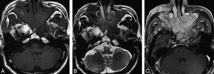
MR imaging of a mass in the nasal cavity and both maxillary sinuses. A, T1-weighted image shows a mass of intermediate intensity compared with muscle and high-intensity areas in the right maxillary sinus. B, Slight hyperintensity is seen on the T2-weighted image. C, Fat-suppressed T1-weighted image has homogeneous gadolinium contrast enhancement.
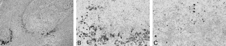
Histopathologic evaluation of the biopsy. A, Hematoxylin-eosin stain reveals lymphoid follicles with a germinal center, among proliferative fibrosis with lymphocyte and plasma cells lacking atypia (original magnification ×25). B, Immunohistopathologic staining for CD138 demonstrates plasma cell infiltration (original magnification ×50). C, Immunostaining demonstrates plasma cell IgG4 expression (original magnification ×50).
References
-
- Ishida M, Hotta M, Kushima R, et al. Multiple IgG4-related sclerosing lesions in the maxillary sinus, parotid gland and nasal septum. Pathol Int 2009; 59: 670–75 - PubMed
-
- Ikeda R, Awataguchi T, Shoji F, et al. A case of paranasal sinus lesions in IgG4-related sclerosing disease. Otolaryngol Head Neck Surg 2010; 142: 458–59 - PubMed
-
- Isaka Y, Yoshioka K, Nishio M, et al. A case of IgG4-related multifocal fibrosclerosis complicated by central diabetes insipidus. Endocr J 2008; 55: 723–28 - PubMed
-
- Boo H, Hogg JP. Nasal cavity neoplasms: a pictorial review. Curr Probl Diagn Radiol 2010; 39: 54–61 - PubMed
-
- Yousem DM, Li C, Montone KT, et al. Primary malignant melanoma of the sinonasal cavity: MR imaging evaluation. Radiographics 1996; 16: 1101–10 - PubMed
Publication types
MeSH terms
Substances
LinkOut - more resources
Full Text Sources
Medical