. 2011 Aug;39(15):6692-704.
doi: 10.1093/nar/gkr252. Epub 2011 May 5.

SHAPE analysis of the FIV Leader RNA reveals a structural switch potentially controlling viral packaging and genome dimerization

Affiliations

SHAPE analysis of the FIV Leader RNA reveals a structural switch potentially controlling viral packaging and genome dimerization

Julia C Kenyon et al. Nucleic Acids Res. 2011 Aug.

Abstract

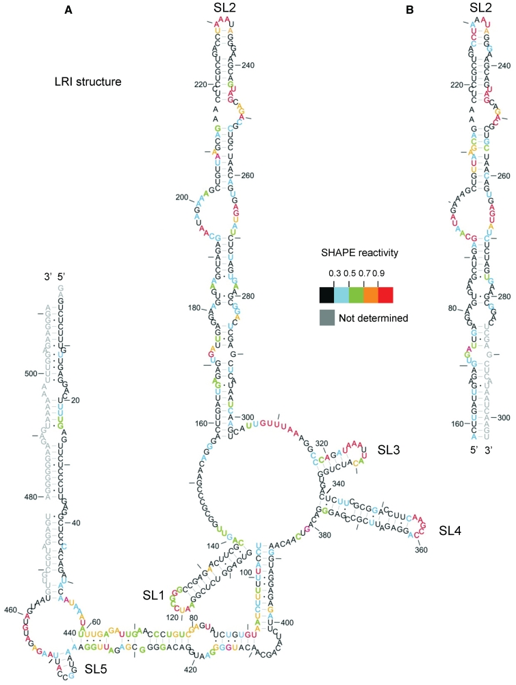
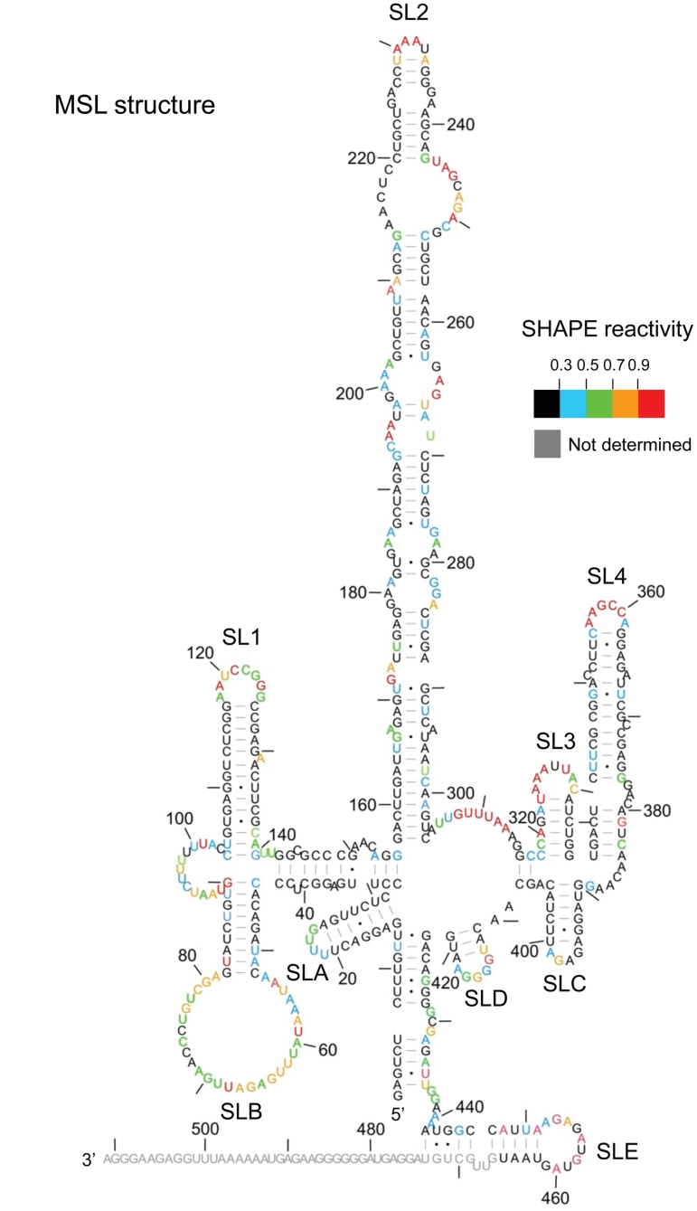
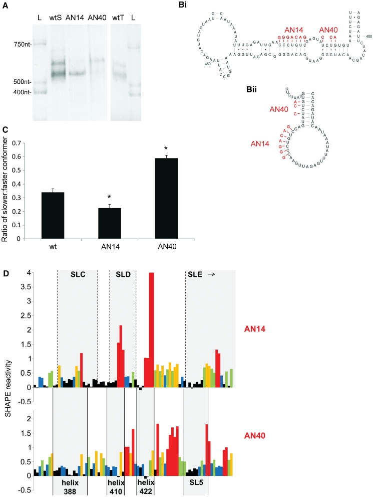
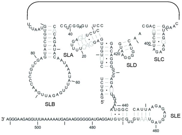
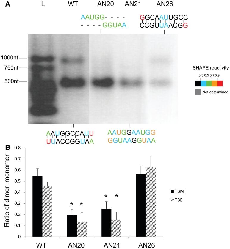
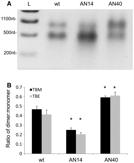
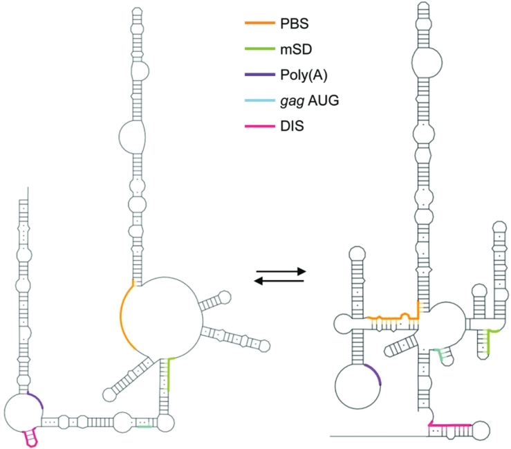
Feline immunodeficiency virus (FIV) infects many species of cat, and is related to HIV, causing a similar pathology. High-throughput selective 2' hydroxyl acylation analysed by primer extension (SHAPE), a technique that allows structural interrogation at each nucleotide, was used to map the secondary structure of the FIV packaging signal RNA. Previous studies of this RNA showed four conserved stem-loops, extensive long-range interactions (LRIs) and a small, palindromic stem-loop (SL5) within the gag open reading frame (ORF) that may act as a dimerization initiation site (DIS), enabling the virus to package two copies of its genome. Our analyses of wild-type (wt) and mutant RNAs suggest that although the four conserved stem-loops are static structures, the 5' and 3' regions previously shown to form LRI also adopt an alternative, yet similarly conserved conformation, in which the putative DIS is occluded, and which may thus favour translational and splicing functions over encapsidation. SHAPE and in vitro dimerization assays were used to examine SL5 mutants. Dimerization contacts appear to be made between palindromic loop sequences in SL5. As this stem-loop is located within the gag ORF, recognition of a dimeric RNA provides a possible mechanism for the specific packaging of genomic over spliced viral RNAs.


Figures

Figure 1.
Figure 1.
SHAPE analysis of the FIV packaging signal RNA. In vitro transcribed RNA was modified with 1M7, reverse transcribed using fluorophore-labelled primers and separated by capillary electrophoresis. Nucleotide positions were determined using G and U sequencing ladders. 1M7 reactivity at each nucleotide position was determined by subtraction of the reverse transcription product of unmodified RNA from that of 1M7-modified RNA, using SHAPEfinder software. Nucleotide reactivities are colour-coded as shown in the key. Reactivities of the 3′-end were not determined (shown in grey). The RNA is drawn in our previously published secondary structure, to ascertain its validity. Nts are numbered with a dash every 10 nt. (A) In vitro transcribed packaging signal RNA; 511 nt. (B) In vitro transcribed SL2 RNA. Data shown are an average of at least two independent experiments.
Figure 2.
Figure 2.
Alternative (MSL) structure of the FIV packaging signal. 1M7 reactivities determined as before were used as pseudo free-energy constraints in a minimal free energy folding algorithm (RNAstructure). Nucleotide reactivities are colour-coded as shown in the key and numbered with a dash every 10 nt.
Figure 3.
Figure 3.
Analyses of the two conformers by non-denaturing PAGE and SHAPE. (A) FIV packaging signal RNA was electrophoresed on a non-denaturing polyacrylamide gel and visualised by ethidium bromide staining and UV illumination. L; RNA ladder wtS; denatured and renatured wt RNA. AN14; denatured and renatured AN14 RNA. AN40; denatured and renatured AN40 RNA. wtT; wt RNA probed after transcription and column purification. (B) Schematic diagram showing the effects of AN14 and AN40 mutations upon: (Bi) the LRI structure; (Bii) SLB of the MSL structure. (C) Densitometric analysis of the ratio of slower-migrating to faster-migrating conformer. Data represent an average of at least six independent experiments. Stars represent statistical significance relative to wt (P < 0.01) by t-test. Error bars show the S.E.M. (D) NMIA reactivity profile of the 3′ portion of each mutant RNA from nts 380–460. Upper panel, AN14; lower panel, AN40. Grey boxes indicate the location of various structural features of each mutant. Reactivity >4 is shown as 4. Numbers below the graph represent the number of the initial nucleotide of each helix in the LRI structure. Data shown are an average of three independent experiments.
Figure 4.
Figure 4.
Structural conservation of the 5′- and 3′-ends of the MSL structure. Sequence variation was previously assessed using an alignment of 76 strains of domestic cat FIV. The ability of each strain of FIV to form this MSL structure was determined using this alignment. Grey nucleotides represent those which vary in sequence but maintain the ability to base pair in 100% of isolates. SLs 1–4 are not shown.
Figure 5.
Figure 5.
Dimerization analysis of SL5 mutants. (A) Dimerization assay and 1M7 reactivity of SL5 mutants. In vitro transcribed FIV packaging signal RNAs containing a wt or mutant SL5 sequence were heated to 95°C, snap-cooled, and incubated at 55°C for 4 h in 10 mM Tris–HCl, pH 7, 200 mM NaCl, 4 mM MgCl2 before electrophoresis on an agarose gel in TBM, or SHAPE analysis as described in ‘Materials and Methods’ section. Mutant designations are given above the relevant lanes and their sequences at SL5 are illustrated above or below each lane. L; RNA ladder. Nucleotide colours show 1M7 reactivities as shown on the key. (B) Densitometric analysis of dimerization on TBM (black bars) or TBE gels (striped bars). Stars represent statistical significance relative to wt (P < 0.01) by t-test. Error bars represent the S.E.M.
Figure 6.
Figure 6.
Dimerization of AN40 and AN14 mutants. Wt, AN40 or AN14 RNA underwent dimerization as described in Figure 5, and the percentage of dimers and monomers was visualized by electrophoresis on 1% agarose gels in TBM or TBE. (A) Representative TBM gel. L; RNA ladder. (B) Densitometric analysis of the ratio of dimer to monomer in wt, AN40 or AN14 RNAs in TBM (black bars) or TBE gels (striped bars). Asterisks represent statistical significance relative to wt by t-test (P < 0.01). Error bars represent the SEM.
Figure 7.
Figure 7.
Accessibility of the functional elements in each structure. Schematic drawing showing the accessibility of the DIS, poly(A), major splice donor (mSD), PBS and gag AUG in the original LRI structure (left) and MSL structure (right).

Similar articles

Cited by

References

    1. Olmsted RA, Hirsch VM, Purcell RH, Johnson PR. Nucleotide sequence analysis of feline immunodeficiency virus: genome organization and relationship to other lentiviruses. Proc. Natl Acad. Sci. USA. 1989;86:8088–8092. - PMC - PubMed
    1. Talbott RL, Sparger EE, Lovelace KM, Fitch WM, Pedersen NC, Luciw PA, Elder JH. Nucleotide sequence and genomic organization of feline immunodeficiency virus. Proc. Natl Acad. Sci. USA. 1989;86:5743–5747. - PMC - PubMed
    1. Yamamoto JK, Sparger E, Ho EW, Andersen PR, O'Connor TP, Mandell CP, Lowenstine L, Munn R, Pedersen NC. Pathogenesis of experimentally induced feline immunodeficiency virus infection in cats. Am. J. Vet. Res. 1988;49:1246–1258. - PubMed
    1. Pedersen NC, Ho EW, Brown ML, Yamamoto JK. Isolation of a T-lymphotropic virus from domestic cats with an immunodeficiency-like syndrome. Science. 1987;235:790–793. - PubMed
    1. Siebelink KH, Chu IH, Rimmelzwaan GF, Weijer K, van Herwijnen R, Knell P, Egberink HF, Bosch ML, Osterhaus AD. Feline immunodeficiency virus (FIV) infection in the cat as a model for HIV infection in man: FIV-induced impairment of immune function. AIDS Res. Hum. Retroviruses. 1990;6:1373–1378. - PubMed

Publication types

Substances