. 2011 Jun;22(6):1041-52.
doi: 10.1681/ASN.2010080808. Epub 2011 May 5.

Mitochondria-targeted peptide accelerates ATP recovery and reduces ischemic kidney injury

Affiliations

Mitochondria-targeted peptide accelerates ATP recovery and reduces ischemic kidney injury

Hazel H Szeto et al. J Am Soc Nephrol. 2011 Jun.

Abstract

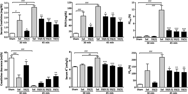
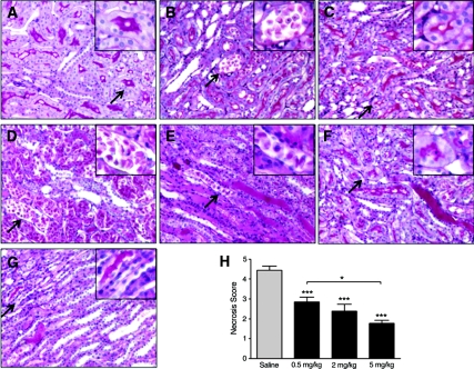
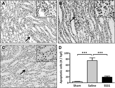
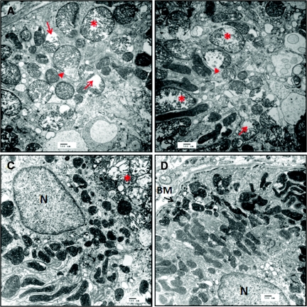
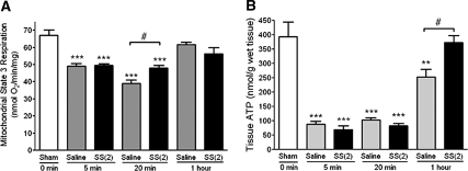
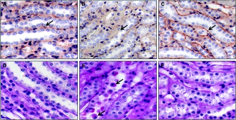
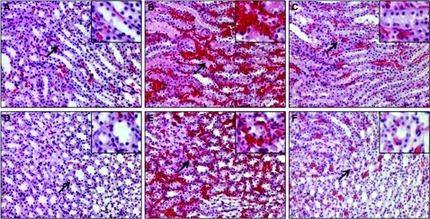
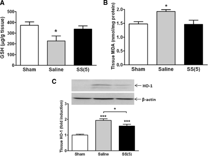
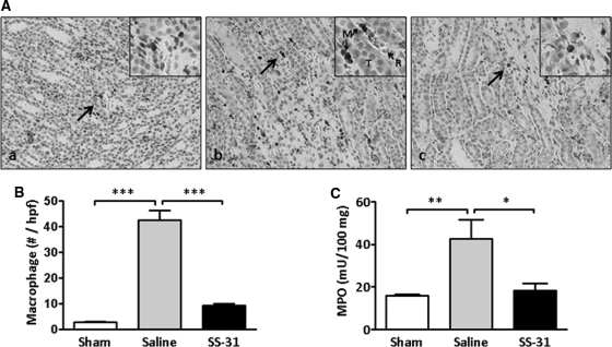
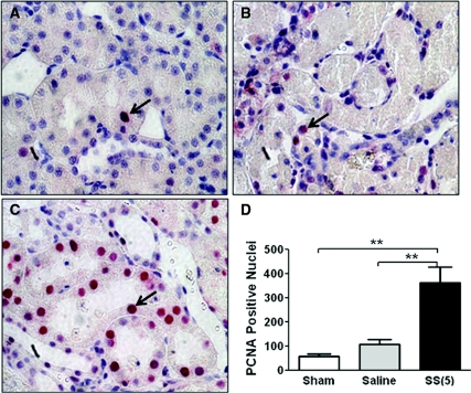
The burst of reactive oxygen species (ROS) during reperfusion of ischemic tissues can trigger the opening of the mitochondrial permeability transition (MPT) pore, resulting in mitochondrial depolarization, decreased ATP synthesis, and increased ROS production. Rapid recovery of ATP upon reperfusion is essential for survival of tubular cells, and inhibition of oxidative damage can limit inflammation. SS-31 is a mitochondria-targeted tetrapeptide that can scavenge mitochondrial ROS and inhibit MPT, suggesting that it may protect against ischemic renal injury. Here, in a rat model of ischemia-reperfusion (IR) injury, treatment with SS-31 protected mitochondrial structure and respiration during early reperfusion, accelerated recovery of ATP, reduced apoptosis and necrosis of tubular cells, and abrogated tubular dysfunction. In addition, SS-31 reduced medullary vascular congestion, decreased IR-mediated oxidative stress and the inflammatory response, and accelerated the proliferation of surviving tubular cells as early as 1 day after reperfusion. In summary, these results support MPT as an upstream target for pharmacologic intervention in IR injury and support early protection of mitochondrial function as a therapeutic maneuver to prevent tubular apoptosis and necrosis, reduce oxidative stress, and reduce inflammation. SS-31 holds promise for the prevention and treatment of acute kidney injury.


Figures

Figure 1.
Figure 1.
SS-31 reduced AKI 24 hours after IR injury. Rats were subcutaneously treated with saline or SS-31 (0.5, 2, or 5 mg/kg) 30 minutes before bilateral occlusion of renal blood flow for 30 or 45 minutes. Treatment was repeated just before onset of reperfusion and at 2 hours after reperfusion. Significant differences were found in all biomarkers among the treatment groups (ANOVA, P < 0.0001). Data are presented as mean ± SEM; n = 6 to 14 in each group. #P < 0.05 and ###P < 0.001 compared with Sham-operated control. *P < 0.05, **P < 0.01, and ***P < 0.001 compared with corresponding saline group.
Figure 2.
Figure 2.
SS-31 reduced tubular injury after ischemia. Rats were subcutaneously treated with saline or SS-31 (0.5, 2 or 5 mg/kg) 30 minutes before bilateral occlusion of renal blood flow for 30 or 45 minutes. Treatment was repeated just before onset of reperfusion and at 2 hours after reperfusion. Kidney sections were stained with PAS. Representative sections from the outer medulla are shown in panels A through G, Magnification: ×200. Inset shows higher magnification of area indicated by the arrow. (A) Kidneys from Sham-operated rats showed normal architecture in the OSOM with prominent brush borders in the proximal tubules (see inset). (B) Focal necrosis was found in the OSOM of saline-treated animals after 30-minute ischemia (inset shows cell sloughing). (C) Necrosis was rare in the SS-31-treated animals after 30-minute ischemia, and brush borders were preserved in many proximal tubules (see inset). (D) Extensive tubular necrosis was found in the OSOM of saline-treated animals after 45-minute ischemia. (E) Architecture of the ISOM was greatly distorted in saline-treated animals after 45 minutes ischemia, and tubules were filled with hyaline casts and sloughed cells. (F) Only focal necrotic areas were observed in the OSOM of SS-31-treated animals after 45-minute ischemia. (G) Architecture of the ISOM was greatly protected by SS-31, with few sloughed cells and occasional casts after 45-minute ischemia. (H) Cell necrosis score after 45-minute ischemia showing dose-dependent protection provided by SS-31 treatment. n = 12 to 14 in each group. ANOVA revealed significant differences among the four groups (P < 0.0001). ***P < 0.001 compared with saline; *P < 0.05 between groups as indicated.
Figure 3.
Figure 3.
SS-31 reduced tubular apoptosis after 30-minute ischemia. Rats were subcutaneously treated with saline or SS-31 (2 mg/kg) 30 minutes before bilateral occlusion of renal blood flow for 30 minutes. Treatment was repeated just before onset of reperfusion and at 2 hours after reperfusion. Apoptotic cells were determined after 24-hour reperfusion by TUNEL stain (brown nuclei). Representative sections from the outer medulla are shown for (A) Sham, (B) saline-treated IR, and (C) SS-31-treated IR rats (magnification: ×200). Inset shows higher magnification of the area indicated by the arrow. (D) Total number of TUNEL-positive cells per high-power field. ***P < 0.001.
Figure 4.
Figure 4.
SS-31 protects kidney mitochondrial structure after IR injury. Rats were subcutaneously treated with saline or SS-31 (2 mg/kg) 30 minutes before occlusion of renal blood flow for 45 minutes. Treatment was repeated just before onset of reperfusion. Ultrastructural studies of kidney sections obtained after 20-minute reperfusion were carried out using transmission electron microscopy. Representative sections from the outer medulla of a saline-treated rat show disruption of normal tubular cytoarchitecture (A and B). Note many rounded, swollen mitochondria with disrupted cristae architecture (*). Some mitochondria show disruption of membranes and release of matrix materials into the cytosol (arrow head). Other swollen mitochondria show complete loss of cristae (arrow). Some normal elongated mitochondria with dense matrix can be observed (B). Representative sections from the outer medulla of a SS-31-treated rat appear quite normal with numerous mitochondria within membrane infoldings near the basement membrane (BM) (C and D). Although a few swollen mitochondria can be found in some cells (*), most mitochondria were elongated with well preserved cristae structure. All images were magnified ×15,000. Scale bar = 500 nm.
Figure 5.
Figure 5.
SS-31 improves kidney mitochondrial function after IR injury. Rats were subcutaneously treated with saline or SS-31 (2 mg/kg) 30 minutes before occlusion of renal blood flow for 45 minutes. Treatment was repeated just before onset of reperfusion. Mitochondria were freshly isolated from kidney tissue obtained at 5, 20, or 60 minutes after reperfusion. ATP was determined from whole kidney tissue obtained at the same time points. (A) Mitochondrial respiration was measured by oxygen consumption using isolated mitochondria with complex I substrates (glutamate/malate) and ADP was added to initiate state 3 respiration. (B) Whole kidney tissue ATP content was determined using the luciferase assay. n = 4 to 8 in each group. **P < 0.01 and ***P < 0.001 compared with Sham; #P < 0.05 comparing saline and SS-31 treatment.
Figure 6.
Figure 6.
SS-31 minimizes renal tubular cell detachment at 1 hour after IR injury. Rats were subcutaneously treated with saline or SS-31 (2 mg/kg) 30 minutes before occlusion of renal blood flow for 45 minutes. Treatment was repeated just before onset of reperfusion, and kidneys were sectioned and stained for β1-integrin by immunohistochemisty and for cell detachment using PAS stain. Panels a through c show β1-integrin staining (reddish-brown stain as shown by arrow) in (A) Sham, (B) saline-treated IR, and (C) SS-31-treated IR rats. β1-integrin was clearly localized to the basal membrane of the tubular cell in Sham and SS-31-treated IR animals, whereas it was diffused throughout the cell in saline-treated IR animals. Panels D through F show that tubular cells are attached to the basement membrane in (D) Sham and (F) SS-31-treated IR animals, but detached cells, some viable (shown by arrow), were clearly found within the tubular lumen of (E) saline-treated IR animals. Magnification: ×600.
Figure 7.
Figure 7.
SS-31 reduces renal medullary vascular congestion 1 hour after IR injury. Rats were subcutaneously treated with saline or SS-31 (2 mg/kg) 30 minutes before occlusion of renal blood flow for 45 minutes. Treatment was repeated just before onset of reperfusion, and kidney sections were stained with hemotoxylin and eosin. Representative sections from the ISOM are shown in panels A through C, and representative sections from the inner medulla are shown in panels D through F (magnification: ×200). Inset shows higher magnification of the area identified by the arrow. Kidneys from Sham-operated rats showed minimal erythrocyte trapping in ISOM and the inner medulla (A, D). Extensive trapping of erythrocytes was found in ISOM and the inner medulla in the saline-treated IR group (B, E). Erythrocyte trapping was greatly reduced in the SS-31-treated IR group (C, F).
Figure 8.
Figure 8.
SS-31 reduces renal oxidative stress caused by IR. Rats were subcutaneously treated with saline or SS-31 (2 mg/kg) 30 minutes before occlusion of renal blood flow for 45 minutes. (A) Effects of SS-31 on renal GSH level after 1-hour reperfusion. n = 6 to 8 in each group. ANOVA, P < 0.05. *P < 0.05 compared with Sham. GSH level in the SS-31-treated IR animals was not different from Sham. (B) Effects of SS-31 on lipid hydroperoxides after 24-hour reperfusion as measured by MDA in renal medullary tissues. n = 3 to 4 in each group. ANOVA, P = 0.01. *P < 0.05 compared with Sham-operated animals. (C) Effects of SS-31 on HO-1 expression at 24 hours after IR injury. Representative western blot for HO-1 from Sham, saline IR, and SS-31 IR animals is shown with β-actin as a loading control. n = 11 to 12 in each group. ANOVA, P < 0.0001. HO-1 protein expression was significantly increased in saline and SS-31 IR animals (***P < 0.001 compared with Sham); however, HO-1 expression in SS-31 animals was significantly lower than saline (*P < 0.05).
Figure 9.
Figure 9.
SS-31 reduces inflammation response to IR injury. (A) Macrophage infiltration in medullary region of (a) Sham, (b) saline-treated IR, and (c) SS-31-treated IR rats. Rats were subcutaneously treated with saline or SS-31 (2 mg/kg) 30 minutes before occlusion of renal blood flow for 30 minutes. Treatment was repeated just before onset of reperfusion and 2 hours later. Kidney sections obtained after 24-hour reperfusion were stained for macrophage using anti-CD68 antibody (brown stain); magnification: ×200. Inset shows area indicated by arrow at higher magnification. Note the presence of macrophages (M) and erythrocytes (R) in capillaries adjacent to tubular cells (T) in the saline IR group. (B) The number of macrophages per high power field were few in the Sham but significantly increased in the saline-treated IR group. SS-31 treatment completely prevented macrophage infiltration. n = 4 to 6 in each group. ANOVA P < 0.0001. ***P < 0.001 between groups. (C) Effects of SS-31 on neutrophil activation 24 hours after 45-minute ischemia as measured by the release of MPO. n = 11 to 13 in each group. ANOVA, P = 0.005, *P < 0.05 and **P < 0.01 between groups.
Figure 10.
Figure 10.
SS-31 accelerates proximal tubular cell regeneration at 24 hours after IR injury. Rats were subcutaneously treated with saline or SS-31 (2 mg/kg) 30 minutes before occlusion of renal blood flow for 45 minutes. Treatment was repeated just before onset of reperfusion and 2 hours later. Representative kidney sections showing immunostaining of PCNA from (A) Sham, (B) saline-treated IR, and (C) SS-31-treated IR animals. Sections were developed using alkaline phosphatase staining, and PCNA+ cells are shown as red nuclei (arrows). PCNA+ cells were very few in the Sham animals and slightly increased 24 hours after IR injury in the saline treatment group, but they were significantly increased in the SS-31 treatment group (D). n = 4 in each group. **P < 0.01 between groups.

Comment in

Similar articles

Cited by

References

    1. Devarajan P: Update on mechanisms of ischemic acute kidney injury. J Am Soc Nephrol 17: 1503–1520, 2006 - PubMed
    1. Brezis M, Rosen S, Silva P, Epstein FH: Selective vulnerability of the medullary thick ascending limb to anoxia in the isolated perfused rat kidney. J Clin Invest 73: 182–190, 1984 - PMC - PubMed
    1. Wirthensohn G, Guder WG: Renal substrate metabolism. Physiol Rev 66: 469–497, 1986 - PubMed
    1. Klein KL, Wang MS, Torikai S, Davidson WD, Kurokawa K: Substrate oxidation by isolated single nephron segments of the rat. Kidney Int 20: 29–35, 1981 - PubMed
    1. Uchida S, Endou H: Substrate specificity to maintain cellular ATP along the mouse nephron. Am J Physiol 255: F977–F983, 1988 - PubMed

Publication types

MeSH terms

Substances