. 2011 Jun;22(6):1053-63.
doi: 10.1681/ASN.2010080841. Epub 2011 May 5.

The pro-apoptotic protein Bim is a microRNA target in kidney progenitors

Affiliations

The pro-apoptotic protein Bim is a microRNA target in kidney progenitors

Jacqueline Ho et al. J Am Soc Nephrol. 2011 Jun.

Abstract

Understanding the mechanisms that regulate nephron progenitors during kidney development should aid development of therapies for renal failure. MicroRNAs, which modulate gene expression through post-transcriptional repression of specific target mRNAs, contribute to the differentiation of stem cells, but their role in nephrogenesis is incompletely understood. Here, we found that the loss of miRNAs in nephron progenitors results in a premature depletion of this population during kidney development. Increased apoptosis and expression of the pro-apoptotic protein Bim accompanied this depletion. Profiling of miRNA expression during nephrogenesis identified several highly expressed miRNAs (miR-10a, miR-106b, miR-17-5p) in nephron progenitors that are either known or predicted to target Bim. We propose that modulation of apoptosis by miRNAs may determine congenital nephron endowment. Furthermore, our data implicate the pro-apoptotic protein Bim as a miRNA target in nephron progenitors.


Figures
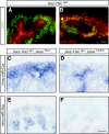
Figure 1.
Figure 1.
Loss of mature miRNAs occurs in Six2-TGCtg/+; DicerFlx/Flx kidneys. Visualization of GFP in Six2-TGCtg/+ E14.5 kidneys co-labeled with pan-cytokeratin (A) or Wt1 (B) showed expression of the fusion GFP-cre protein in nephron progenitors. Scale bar, 20 μm. LNA-ISH for mmu-miR-10a or mmu-miR-30a in E14.5 Six2-TGCtg/+; DicerFlx/+ (C and E) and Six2-TGCtg/+; DicerFlx/Flx (D and F) kidneys showed the loss of miRNA expression in nephron progenitors and their derivatives in Six2-TGCtg/+; DicerFlx/Flx mutant kidneys. Scale bar, 50 μm.
Figure 2.
Figure 2.
Premature depletion of nephron progenitors in Six2-TGCtg/+; DicerFlx/Flx kidneys during nephrogenesis. Histologic analysis of Six2-TGCtg/+; DicerFlx/+ (A and C) and Six2-TGCtg/+; DicerFlx/Flx (B and D) E14.5 kidney sections and E16.5 showed a marked loss of nephron progenitors in E16.5 mutant kidneys. In each panel, the double-headed arrows delimit the extent of progenitors. Scale bar, 20 μm. Lineage tracing with the Rosa26-lacZ reporter allele showed that β-galactosidase expression remains confined to the nephron lineage in Six2-TGCtg/+; DicerFlx/Flx kidneys (F and H) and is greatly reduced compared with control kidneys (E and G). Scale bar, 50 μm.
Figure 3.
Figure 3.
ISH for known transcription factors in nephron progenitors for E16.5 Six2-TGCtg/+; DicerFlx/+ (A, E, I, M, Q, and U) and Six2-TGCtg/+; DicerFlx/Flx (B, F, J, N, R, and V) kidneys reveals decreased expression of Six2, Eya1, Osr1, Wt1, Pax2, and Sall1 in the mutant kidneys. The stromal markers FoxD1 and Pod1 (G–L) remained unchanged in their expression pattern, and a small decrease in Raldh2 expression (C and D) in the cortical stroma of E16.5 Six2-TGCtg/+; DicerFlx/Flx was observed. The expression of the ureteric bud marker, c-ret (O and P), was unchanged. Wnt4 expression was present in the pretubular aggregates of mutant Six2-TGCtg/+; DicerFlx/Flx kidneys (S and T). Scale bar, 50 μm. CM, cap mesenchyme; SM, stromal mesenchyme; UB, ureteric bud; PA, pretubular aggregate.
Figure 4.
Figure 4.
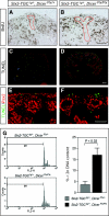
Increased apoptosis in the nephrogenic zone occurs in Six2-TGCtg/+; DicerFlx/Flx kidneys. Proliferating cells in E15.5 kidneys were identified by BrdU uptake. The number of BrdU+ nephron progenitors was similar between Six2-TGCtg/+; DicerFlx/+ (A) and Six2-TGCtg/+; DicerFlx/Flx (B) kidneys. The ureteric bud is outlined in red. Scale bar, 50 μm. Increased TUNEL+ (green) cells occurred in the nephrogenic zone of Six2-TGCtg/+; DicerFlx/Flx (D and F) compared with Six2-TGCtg/+; DicerFlx/+ (C and E) kidneys. Co-labeling with Pax2 (red) marked the ureteric bud and metanephric mesenchyme. Scale bars: 100 (C and D) and 50 μm (E and F). (G) (Left) Representative DNA content histogram plots for E15.5 Six2-TGCtg/+; DicerFlx/+ and Six2-TGCtg/+; DicerFlx/Flx nephron progenitors, as determined by propidium iodide staining for DNA content and subsequent dual-color flow cytometric analysis gating on GFP+ nephron progenitors. The M1 gate denotes DNA content <2n. (Right) Frequency (%) of <2n DNA-containing components (i.e., apoptotic cells or cellular fragments) in Six2-TGCtg/+; DicerFlx/+ and Six2-TGCtg/+; DicerFlx/Flx nephron progenitors, determined by propidium iodide staining and dual-color flow cytometry (mean ± SE, for three independent experiments). There was a significant increase in the frequency of cells with <2n content in Six2-TGCtg/+; DicerFlx/Flx nephron progenitors (17 ± 3.1%) compared with Six2-TGCtg/+; DicerFlx/+ nephron progenitors (3.6 ± 1.5%; P < 0.02; t test; n = 10 embryos for each of three independent experiments).
Figure 5.
Figure 5.
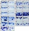
miRNA expression profiled in the embryonic kidney. (A) Northern blots for mmu-miR-30a, mmu-miR-106b, mmu-miR-130a, and mmu-miR-335 in a panel of wild-type adult and embryonic tissues confirmed the presence of these miRNAs in the embryonic kidney. 5S rRNA, loading control. (B) Expression of mmu-let-7c, mmu-miR-10a, mmu-miR-17-5p, mmu-miR-23b, mmu-miR-24, mmu-miR-26a, mmu-miR-30a, mmu-miR-30c, mmu-miR-106b, mmu-miR130a, and mmu-miR-335 in E14.5 wild-type CD-1 kidneys by LNA-ISH. Note an air bubble in the mmu-miR-10a figure. Scrambled oligonucleotide, negative control. Scale bar, 50 μm.
Figure 6.
Figure 6.
The expression of the candidate miRNA target, Bim, is increased in Six2-TGCtg/+; DicerFlx/Flx mutant kidneys. (A) Sequence alignment of the Bim 3′UTR and mmu-miR-10a, mmu-miR-17-5p, and mmu-miR-106b with two to three conserved target site matches for each miRNA (nucleotides in blue possess Watson-Crick pairing between the miRNA and Bim 3′UTR, whereas nucleotides in red are unpaired). (B) Western blot analysis showed an increase in the BimEL isoform in Six2-TGCtg/+; DicerFlx/Flx (M) kidneys compared with control kidneys (C) at E15.5 and E16.5. The BimL and BimS isoforms were relatively unchanged, as were Bcl2 and GAPDH. (C) Immunohistochemical staining for Bim and Bcl2 showed increased Bim levels specifically in nephron progenitors in mutant Six2-TGCtg/+; DicerFlx/Flx kidneys, whereas Bcl2 expression was constant. Scale bar, 20 μm.

Similar articles

Cited by

References

    1. Dressler GR: The cellular basis of kidney development. Annu Rev Cell Dev Biol 22: 509–529, 2006 - PubMed
    1. Schedl A: Renal abnormalities and their developmental origin. Nat Rev Genet 8: 791–802, 2007 - PubMed
    1. Bernstein E, Kim SY, Carmell MA, Murchison EP, Alcorn H, Li MZ, Mills AA, Elledge SJ, Anderson KV, Hannon GJ: Dicer is essential for mouse development. Nat Genet 35: 215–217, 2003 - PubMed
    1. Kanellopoulou C, Muljo SA, Kung AL, Ganesan S, Drapkin R, Jenuwein T, Livingston DM, Rajewsky K: Dicer-deficient mouse embryonic stem cells are defective in differentiation and centromeric silencing. Genes Dev 19: 489–501, 2005 - PMC - PubMed
    1. Wang Y, Medvid R, Melton C, Jaenisch R, Blelloch R: DGCR8 is essential for microRNA biogenesis and silencing of embryonic stem cell self-renewal. Nat Genet 39: 380–385, 2007 - PMC - PubMed

Publication types

MeSH terms