. 2011 Aug;301(2):G385-400.
doi: 10.1152/ajpgi.00430.2010. Epub 2011 May 5.

cAMP-guanine exchange factor protection from bile acid-induced hepatocyte apoptosis involves glycogen synthase kinase regulation of c-Jun NH2-terminal kinase

Affiliations

cAMP-guanine exchange factor protection from bile acid-induced hepatocyte apoptosis involves glycogen synthase kinase regulation of c-Jun NH2-terminal kinase

A Johnston et al. Am J Physiol Gastrointest Liver Physiol. 2011 Aug.

Abstract

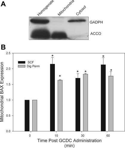
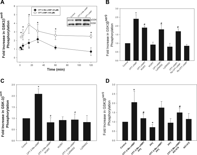
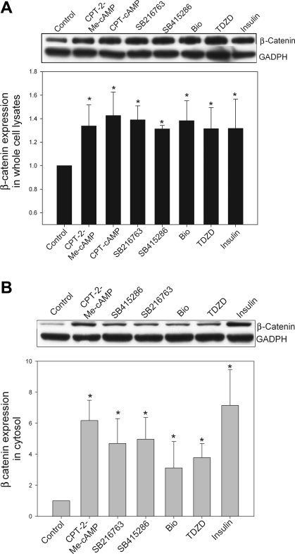
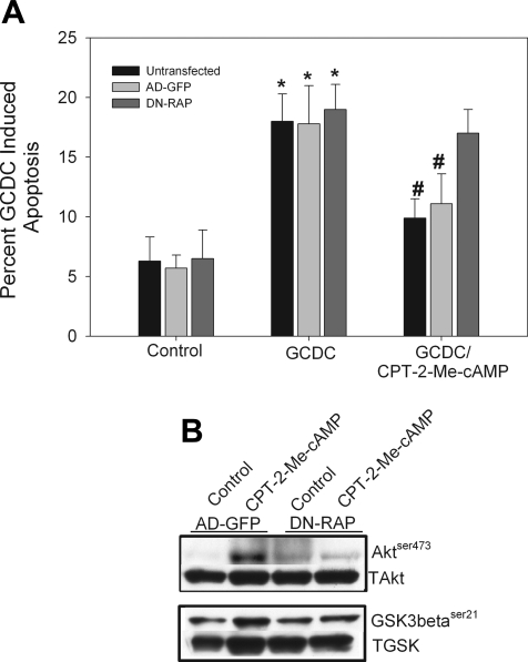
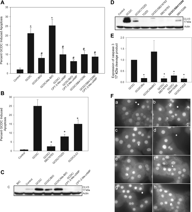
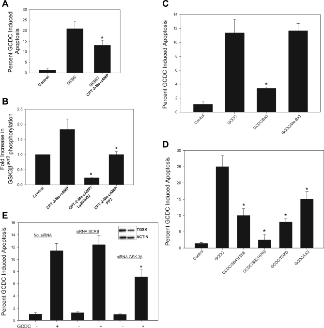
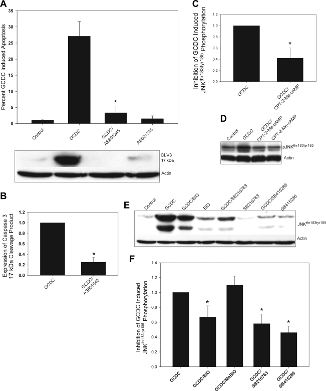
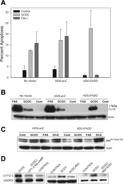
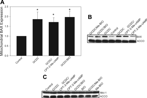
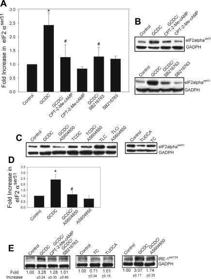
Cholestatic liver disorders are accompanied by the hepatic accumulation of cytotoxic bile acids that induce cell death. Increases in cAMP protect hepatocytes from bile acid-induced apoptosis by a cAMP-guanine exchange factor (cAMP-GEF)/phosphoinositide-3-kinase (PI3K)/Akt pathway. The aim of these studies was to identify the downstream substrate in this pathway and to determine at what level in the apoptotic cascade cytoprotection occurs. Since inhibitory phosphorylation of glycogen synthase kinase-3 (GSK) occurs downstream of PI3K/Akt and this phosphorylation has been implicated in cell survival, we conducted studies to determine whether GSK was downstream in cAMP-GEF/PI3K/Akt-mediated cytoprotection. Our results show that treatment of hepatocytes with the cAMP-GEF-specific analog, 4-(4-chlorophenylthio)-2'-O-methyladenosine-3',5'-cAMP, results in PI3K-dependent phosphorylation of GSK. Direct chemical inhibition of GSK in rat hepatocytes or human HUH7-NTCP cells with several structurally and functionally distinct inhibitors including bromoindirubin-3'-oxime (BIO), maleimides (SB216763, SB415286), thiadiazolidine derivatives, and LiCl attenuates apoptosis induced by glycochenodeoxycholate (GCDC). In addition, genetic silencing of the GSK β isoform with small interfering RNA attenuates GCDC apoptosis in HUH7-NTCP cells. Adenoviral inhibition of the Rap1 blocks both cAMP-GEF-mediated cytoprotection against GCDC-induced apoptosis and Akt/GSK3β phosphorylation. GCDC-induced phosphorylation of the proapoptotic kinase, c-Jun NH(2)-terminal kinase (JNK) is inhibited by GSK inhibition or cAMP-GEF activation. GCDC-induced apoptosis is accompanied by phosphorylation of the endoplasmic reticulum stress markers pIEF2α and IRE-1, and pretreatment with the cAMP-GEF analog or GSK inhibitors prevents this phosphorylation. Collectively, our results support the presence of a cAMP/cAMP-GEF/Rap1/PI3K/Akt/GSKβ survival pathway in hepatocytes that inhibits bile acid-induced JNK phosphorylation.


Figures

Fig. 1.
Fig. 1.
Mitochondrial fractions prepared from subcellular fractionization and digitonin permeabilization are equivalent. A: rat hepatocytes were solubilized with digitonin as indicated in materials and methods to isolate a digitonin-soluble fraction (called cytosol) and a digitonin-resistant fraction that was subsequently solubilized in Triton X-100 (mitochondria). Equal aliquots of each preparation were immunoblotted with the cytosolic marker protein GADPH and the mitochondrial marker cytochrome c oxidase (ACCO). B: rat hepatocytes were treated with glycochenodeoxycholate (GCDC) for the indicated times and then a mitochondrial fraction was prepared by either subcellular fractionization (SCF) or selective digitonin permeabilization (Dig Perm). Then the amount of the proapoptotic protein, BAX, in each mitochondrial fraction was determined by immunoblotting. *Amount is significantly different than that in control untreated cultures.
Fig. 2.
Fig. 2.
Phosphorylation of glycogen synthase kinase (GSK)-3 (GSK3β) by cAMP analogs in rat hepatocytes. A: whole cell lysates were prepared from rat hepatocytes treated with 8-(4-chlorophenylthio)-cAMP (CPT-cAMP; 100 μM) or 4-(4-chlorophenylthio)-2′-O-methyladenosine-3′,5′-cyclic monophosphate (CPT-2-Me-cAMP; 20 μM) for the indicated time period, and the amount of GSK3βser9 phosphorylation was determined by immunoblotting with phospho-specific antibodies. Equal protein loading was verified by reprobing for total GSK3β expression. Results are expressed as the fold increase in the amount of GSK3βser9 phosphorylation compared with untreated controls. Inset: representative blot of the amount of phosphorylated (pGSK) and total GSK3β (tGSK) in hepatocytes cultures treated with the indicated concentration of the analogs for 30 min. *Significantly different than the amount in untreated control cultures. B: downstream mediators of cAMP-induced phosphorylation of GSK3βser9 were determined by immunoblotting of cell lysates prepared from cultures treated with CPT-cAMP for 15 min in the presence and absence of phosphoinositide-3-kinase (PI3K) inhibitors wortmannin (WOT; 50 nM) or Ly294002 (20 μM) or 500 nM of the protein kinase A inhibitor Rp-8-(4-chlorophenylthio)-cAMP (Rp-CPT-cAMP). *Significantly different than control. #Significantly different from CPT-cAMP-treated hepatocytes. The downstream mediators of cAMP-GEF-induced phosphorylation GSK3βser9 were determined by immunoblotting of cell lysates from cultures treated with CPT-2-Me-cAMP for 15 min in the presence and absence of PI3K inhibitors wortmannin (50 nM) or Ly294002 (20 μM) (C) or epidermal growth factor inhibitor AG1478 (5 μM) or 10 μM of the Src inhibitor 4-amino-5-(4-chlorophenyl)-7-(t-butyl)pyrazolo[3,4-d]pyrimidine (PP2) or its inactive control compound 4-amino-7-phenylpyrazol [3,4-d]pyrimidine (PP3) (D). *Significantly different than control. #Significantly different from CPT-2-Me-cAMP-treated hepatocytes.
Fig. 3.
Fig. 3.
cAMP-GEF activation and GSK inhibition increase β-catenin levels in hepatocytes. Rat hepatocyte cultures were treated for 2 h with CPT-2-Me-cAMP (20 μM), SB415286 (25 μM), SB216763 (10 μM), bromoindirubin-3′-oxime (BIO; 10 μM), 4-benzyl-2-methyl-1,2,4-thiadiazolidine-3,5-dione (TDZD; 40 μM), or insulin (50 nM). Whole cell lysates and cytosol extracts were prepared. Aliquots of each preparation were immunoblotted with antibodies to β-catenin, and equal protein loading was verified with immunoblotting for GADPH. Representative blots are shown in A and B along with graphical depiction of the results of 3 separate experiments. *Significantly different from control cells treated with only vehicle.
Fig. 4.
Fig. 4.
cAMP-GEF-mediated cytoprotection and activation of PI3K/Akt is Rap1 dependent. Rat hepatocytes were transfected with a green fluorescent protein (GFP) adenovirus (AD-GFP) or a Rap1GAP-GFP adenovirus (DN-RAP) for 24 h and then treated with GCDC (100 μM) for 2 h in the presence and absence of 20 μM CPT-2-Me-cAMP. Apoptosis was monitored morphologically in Hoechst-stained cells (A). Rat hepatocytes were transfected with a control GFP adenovirus or a Rap1GAP-GFP adenovirus for 24 h then treated for 15 min with CPT-2-Me-cAMP and the amount of phosphorylated Aktser473 and GSK3βser9 determined and normalized to total Akt (TAkt) or GSK (TGSK). by immunoblotting. B: representative immunoblots from 3 separate experiments are shown.
Fig. 5.
Fig. 5.
Inhibition of GSK attenuates bile acid-induced apoptosis in rat hepatocytes. Hepatocyte cultures were treated with GCDC (50 μM) for 2 h in the presence or absence of the GSK inhibitors SB415286 (25 μM), 10 μM of BIO, or its inactive analog ME-BIO (A) or with TDZD (40 μM) or SB216763 (10 μM) (B). In some experiments 20 μM of CPT-2-Me-cAMP was also added. The amount of apoptosis was determined by morphological examination of Hoechst stained cells (A and B) or biochemical analysis for the presence of the 17-kDa cleavage fragment of caspase (CLV3) (C–E). Equal protein loading was verified by reprobing CLV3 blots with actin. Representative blots are shown in C and D with the quantification of 3 separate experiments in E. The composite photomicrograph of Hoechst-stained cells (F) shows control hepatocytes (a) and hepatocytes treated with GCDC (50 μM) alone (b) or after 30-min pretreatment with CPT-2-Me-cAMP (20 μM, c), SB216763 (5 μM, d), SB415286 (5 μM, e), BIO (5 μM, f), TDZD (40 μM, g), or the JNK inhibitor AS601645 (5 μM, h). Magnification ×100. *Significantly different than untreated control cultures. #Significantly different from GCDC-treated cultures.
Fig. 6.
Fig. 6.
Activation of cAMP-GEF and inhibition of GSK protect against bile acid-induced apoptosis in human hepatoma cells. A: HUH7-NTCP cells were pretreated with CPT-2-Me-cAMP for 30 min and then exposed to GCDC (200 μM) for 3 h. Apoptosis was determined by morphological evaluation of Hoechst-stained cells. B: HUH7-NTCP cells were treated with CPT-2-Me-cAMP for 30 min in the presence or absence of Ly294002 (20 μM) or PP2 (10 μM) and the amount of GSK3βser9 phosphorylation was determined in cell lysates. HUH7-NTCP cells were pretreated with the GSK inhibitor BIO (10 μM) or it inactive control analog ME-BIO (10 μM) (C) or with SB415286 (25 μM), SB216746 (10 μM), or TDZD (25 μM) (D) and then exposed to GCDC (200 μM) for 3 h. Apoptosis was determined by morphological evaluation of Hoechst-stained cells. E: HUH7-NTCP cells were transfected with small interfering RNA (siRNA) to GSK3β or a scrambled siRNA (siRNA SCRB) for 48 h and then exposed to GCDC for 4 h, and the percent apoptosis was determined morphologically. *Significantly different than GCDC (A, C–E)- or CPT-2-Me-cAMP (B)-treated cells.
Fig. 7.
Fig. 7.
GSK inhibition prevents bile acid-induced phosphorylation of JNK. A and B: rat hepatocytes were treated with GCDC (50 μM) for 1 h in the presence or absence the JNK inhibitor. AS601245 (10 μM) and the amount of apoptosis determined by morphological evaluation of Hoechst-stained cells or Western blotting for the active 17-kDa cleaved fragment of caspase 3 (CLV3; A). B–D: quantification of 3 separate experiments. Rat hepatocytes were treated with GCDC (50 μM) for 1 h in the presence or absence of the CPT-2-Me-cAMP (20 μM) and the amount of phosphorylated JNKthr183,tyr185 (pJNKthr183,tyr185) determined by immunoblotting. A representative blot is shown in C and the quantification of immunoblots from 4 separate experiments is shown in D. Results are expressed as the change in the amount of phosphorylated JNK compared with that seen with GCDC. Rat hepatocytes were treated with GCDC (50 μM) for 1 h in the presence and absence of the GSK inhibitors BIO (10 μM), SB216746 (10 μM), or SB415286 (25 μM) and the amount of phosphorylated JNKthr183,tyr185 was determined by immunoblotting. A representative blot is shown in E, and the quantification of immunoblots from 5 separate experiments shown in F. *Significantly different from GCDC-treated hepatocytes.
Fig. 8.
Fig. 8.
Bile acid-induced apoptosis and JNK activation in rat hepatocytes is death receptor independent. Rat hepatocytes were transfected with an adenoviral control AD5LacZ and dominant-negative AD5delta-FADD construct and then treated with GCDC (50 μM), deoxycholate (DCA, 100 μM) for 2 h, or Fas ligand (Fas L; 50 ng/ml) for 4 h. Apoptosis was monitored morphologically by examination of Hoechst-stained cells (A) or biochemically by immunoblotting for CLV3 (B). Cont, control. Equal protein loading was verified by immunoblotting for actin. The amount of phosphorylated JNKthr183,tyr185 was determined by immunoblotting (C). D: rat hepatocytes were pretreated with CPT-2-Me-cAMP (20 μM), BIO (10 μM), or with SB216763 (10 μM) and then exposed to GCDC for 2 h. Cytosolic fractions were prepared by subcellular fractionization as described in materials and methods. The amount of cytochrome c (CYTO C) released into cytosol was determined by immunoblotting. *Significantly different than in the presence of GCDC.
Fig. 9.
Fig. 9.
Cytoprotection by cAMP-GEF activation or GSK inhibition is independent of BAX or Mcl-1. Rat hepatocytes were pretreated with CPT-2-Me-cAMP (20 μM) or the GSK inhibitors BIO (10 μM) or it inactive analog ME-BIO (10 μM) for 30 min and then exposed to GCDC for 60 min. Mitochondrial fractions were prepared by selective digitonin permeabilization as described in materials and methods. Mitochondrial fractions were immunoblotted for BAX (A and B) or Mcl-1 (C). Representative gels are shown in B and C with quantification of the BAX expression in A. Equal protein loading was verified by immunoblotting for the mitochondrial specific protein ACCO. *Significantly different than the amount of protein in control untreated cells.
Fig. 10.
Fig. 10.
Bile acid-induced apoptosis and activation of JNK involves induction of the endoplasmic reticulum stress response. Rat hepatocytes were treated with hydrophobic cytotoxic bile acids GCDC (50 μM), taurochenodeoxycholate (TCDC, 100 μM), or taurolithocholate (TLC, 25 μM) or the hydrophobic cytoprotective bile acids tauroursodeoxycholate (TUDCA, 100 μM) or taurocholate (TC, 100 μM) for 1 h in the presence or absence the 20 μM CPT-2-Me-cAMP, 10 μM SB216763, or 10 μM AS604850, and the amounts of phosphorylated peIF2αser51 (A–D) and phosphorylated IRE-1ser724 (E) were determined by immunoblotting. Equal protein loading was verified by immunoblotting for actin or GADPH. Representative immunoblots are shown in B, C, and E, and the quantification of immunoblots from 3 separate experiments is presented in A and D. *Significantly different from control cells treated; #significantly different from bile acid-treated cells.

References

    1. Aghdam SY, Barger SW. Glycogen synthase kinase-3 in neurodegeneration and neuroprotection: lessons from lithium. Curr Alzheimer Res 4: 21–31, 2007 - PubMed
    1. Avila J, Wandosell F, Hernández F. Role of glycogen synthase kinase-3 in Alzheimer's disease: pathogenesis and glycogen kinase-3 inhibitors. Expert Rev Neurother 10: 703–710, 2010 - PubMed
    1. Bain J, Plater L, Elliott M, Shpiro N, Hastie CJ, McLauchlan H, Klevernic I, Arthur JS, Alessi DR, Cohen P. The selectivity of protein kinase inhibitors: a further update. Biochem J 408: 297–315, 2007 - PMC - PubMed
    1. Baviera AM, Zanon NM, Navegantes LC, Kettelhut IC. Involvement of cAMP/Epac/PI3K-dependent pathway in the antiproteolytic effect of epinephrine on rat skeletal muscle. Mol Cell Endrocrinol 315: 104–112, 2010 - PubMed
    1. Beauchard A, Ferandin Y, Frère S, Lozach O, Blairvacq M, Meijer L, Thiéry V, Besson T. Synthesis of novel 5-substituted indirubins as protein kinases. Bioorg Med Chem 14: 6434–6443, 2006 - PubMed

Publication types

MeSH terms

Substances