. 2011 Jun 24;109(1):80-5.
doi: 10.1161/CIRCRESAHA.110.237594. Epub 2011 May 5.

Pannexin1 regulates α1-adrenergic receptor- mediated vasoconstriction

Affiliations

Pannexin1 regulates α1-adrenergic receptor- mediated vasoconstriction

Marie Billaud et al. Circ Res. .

Abstract

Rationale: The coordination of vascular smooth muscle cell constriction plays an important role in vascular function, such as regulation of blood pressure; however, the mechanism responsible for vascular smooth muscle cell communication is not clear in the resistance vasculature. Pannexins (Panx) are purine-releasing channels permeable to the vasoconstrictor ATP and thus may play a role in the coordination of vascular smooth muscle cell constriction.

Objective: We investigated the role of pannexins in phenylephrine- and KCl-mediated constriction of resistance arteries.

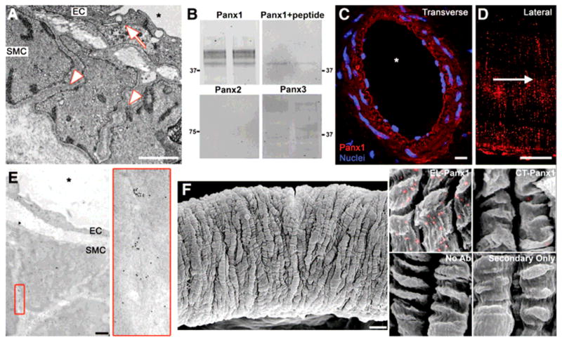
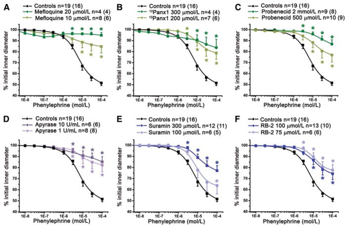
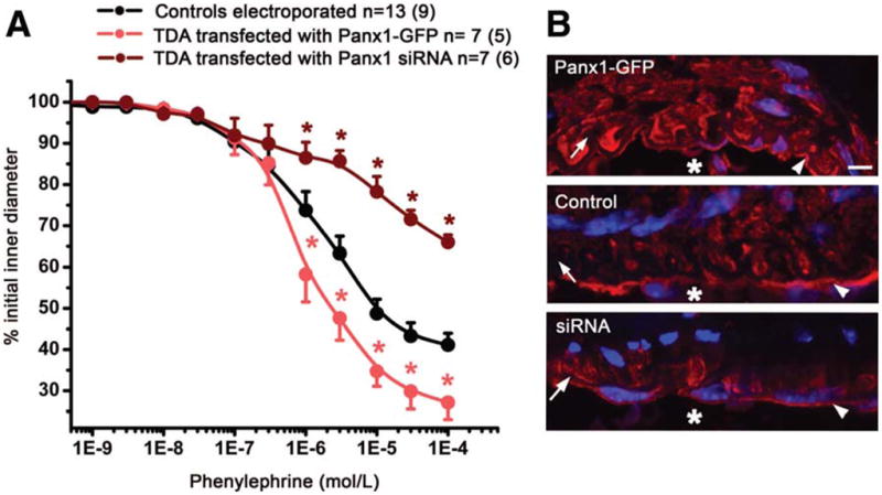
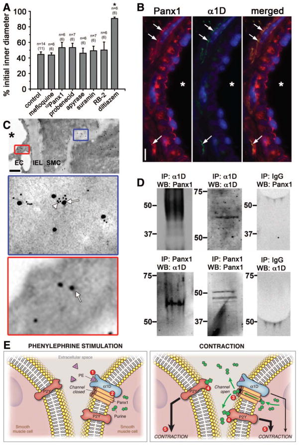
Methods and results: Western blot, immunohistochemistry, and immunogold labeling coupled to scanning and transmission electron microscopy revealed the presence of Panx1 but not Panx2 or Panx3 in thoracodorsal resistance arteries. Functionally, the contractile response of pressurized thoracodorsal resistance arteries to phenylephrine was decreased significantly by multiple Panx inhibitors (mefloquine, probenecid, and (10)Panx1), ectonucleotidase (apyrase), and purinergic receptor inhibitors (suramin and reactive blue-2). Electroporation of thoracodorsal resistance arteries with either Panx1-green fluorescent protein or Panx1 small interfering RNA showed enhanced and decreased constriction, respectively, in response to phenylephrine. Lastly, the Panx inhibitors did not alter constriction in response to KCl. This result is consistent with coimmunoprecipitation experiments from thoracodorsal resistance arteries, which suggested an association between Panx1 and α1D-adrenergic receptor.

Conclusions: Our data demonstrate for the first time a key role for Panx1 in resistance arteries by contributing to the coordination of vascular smooth muscle cell constriction and possibly to the regulation of blood pressure.


Figures

Figure 1
Figure 1. Panx1 is expressed in the membrane of VSMC in TDA
(A) Representative TEM image of TDA demonstrates close apposition (arrow) between EC, whereas VSMC are separated by a large intercellular space (arrowheads). (B) Duplicate western blots of TDA lysates for the three Panx isoforms (Panx1, Panx2 and Panx3) and Panx1 antibody incubated with cognate peptide. (C) immunofluorescence of a transverse section of TDA labeled for Panx1 (red), nuclei are stained with DAPI (blue). (D) Immunofluorescence of a lateral view of VSMC of a TDA labeled for Panx1 (red), arrow indicates direction of blood flow. (E) Immuno-TEM labeling of TDA for Panx1; enlargement of red box is shown on right. (F) Left: representative SEM of a TDA. Right: enlarged images of VSMC from immuno-SEM labeled for Panx1 (gold beads were pseudocolored in pink). Samples were labeled with antibodies against the extracellular loop of Panx1 (EL-Panx1) or the C-terminal of Panx1 (CT-Panx1); VSMC without labeling or primary antibodies are also shown. Scale bar is 1μm in (A) and (E), 5μm in (D), 10μm in (C) and (F). Asterisks indicate vessel lumen.
Figure 2
Figure 2. Panx is involved in PE constriction of TDA
The effect of cumulative concentrations of PE on internal diameter of pressurized TDA was significantly decreased after treatment with Panx inhibitors: (A) mefloquine (10 μmol/L), (B) 10Panx1 peptide (200 μmol/L) and (C) probenecid (2 mmol/L and 500μmol/L). (D), effect of apyrase (1 U/mL and 10 U/mL). We also inhibited purinergic receptors with suramin (100 μmol/L and 300 μmol/L), (E) and P2Y receptors with RB-2 (75 μmol/L); (F). n indicates the number of vessels and the value in parentheses is the number of mice. *P<0.05.
Figure 3
Figure 3. Modulation of Panx1 expression in VSMC modifies PE-induced constriction of TDA
The VSMC of TDA were transfected with either a plasmid containing Panx1-GFP or Panx1 siRNA. (A) The effect of cumulative concentrations of PE was investigated in each condition. (B) Immunofluoresence of TDA transfected with Panx1-GFP (upper panel), untransfected TDA (middle panel) and TDA transfected with Panx1 siRNA (lower panel) labeled for Panx1. Scale bar is 5μm. The asterisks in A indicates P<0.05 and in B, the asterisks indicates vessel lumen. In B, arrows point to IEL, arrowheads point to EC.
Figure 4
Figure 4. Panx1 is associated with α1D -adrenoreceptor and is not involved in KCl constriction
(A) Previously used inhibitors (mefloquine (10 μmol/L); 10Panx1 (200 μmol/L), probenecid (2 mmol/L) apyrase (10 U/mL), suramin (300 μmol/L) or RB-2 (75 μmol/L)) had no effect on 40 mmol/L KCl-induced constriction. However, the L-type Ca2+ channel inhibitor diltiazem (10 μmol/L) significantly inhibited the KCl-induced constriction. *P<0.05. (B) Immunolabeling of transverse sections of TDA labeled for Panx1 (red) and α1D-adrenoreceptor (green). Nuclei are stained in blue and asterisks indicate the lumen. Arrows point to colocalized labeling for Panx1 and α1D-adrenoreceptor. Scale bar is 10μm. (C) Immunogold labeling of TDA for Panx1 (arrows) and α1D-adrenoreceptors (arrowheads). Scale bar is 1 μm. (D) Interaction between Panx1 with α1D-adrenoreceptors was revealed when lysates from TDA were immunoprecipitated (IP) either with α1D-adrenoreceptor (α1D, upper left panel) or Panx1 (lower left panel) and blotted (WB) with Panx1 or α1D-adrenoreceptor antibodies, respectively. Panx1 and α1D-adrenoreceptor pull down were verified by blotting with Panx1 or α1D-adrenoreceptor antibodies (middle panels) and the absence of IgG was also controlled (right panels). (E) Summary of PE-induced vasoconstriction: application of PE stimulates α1D-adrenoreceptor (1) followed by the activation of Panx1 channel (2) leading to the opening of the Panx1 channel and the release of purines in the extracellular space (3). Purines bind to P2Y receptors (4) reinforcing the α1D-adrenoreceptor constriction (5).

References

    1. Segal SS, Duling BR. Flow control among microvessels coordinated by intercellular conduction. Science. 1986;234:868–870. - PubMed
    1. Fanchaouy M, Serir K, Meister JJ, Beny JL, Bychkov R. Intercellular communication: role of gap junctions in establishing the pattern of ATP-elicited Ca2+ oscillations and Ca2+-dependent currents in freshly isolated aortic smooth muscle cells. Cell Calcium. 2005;37:25–34. - PubMed
    1. Siegl D, Koeppen M, Wolfle SE, Pohl U, de Wit C. Myoendothelial coupling is not prominent in arterioles within the mouse cremaster microcirculation in vivo. Circ Res. 2005;97:781–788. - PubMed
    1. Hakim CH, Jackson WF, Segal SS. Connexin isoform expression in smooth muscle cells and endothelial cells of hamster cheek pouch arterioles and retractor feed arteries. Microcirculation. 2008;15:503–514. - PMC - PubMed
    1. D'Hondt C, Ponsaerts R, De Smedt H, Bultynck G, Himpens B. Pannexins, distant relatives of the connexin family with specific cellular functions? Bioessays. 2009;31:953–974. - PubMed

Publication types

MeSH terms