. 2011 May-Jun;5(3):237-48.
doi: 10.4161/cam.5.3.15909. Epub 2011 May 1.

GIV/Girdin is a rheostat that fine-tunes growth factor signals during tumor progression

Affiliations

GIV/Girdin is a rheostat that fine-tunes growth factor signals during tumor progression

Pradipta Ghosh et al. Cell Adh Migr. 2011 May-Jun.

Abstract

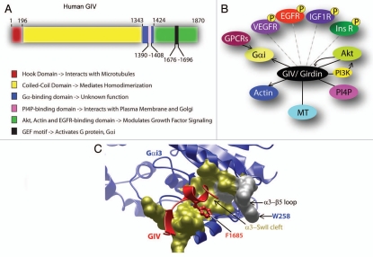
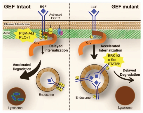
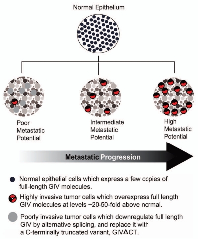
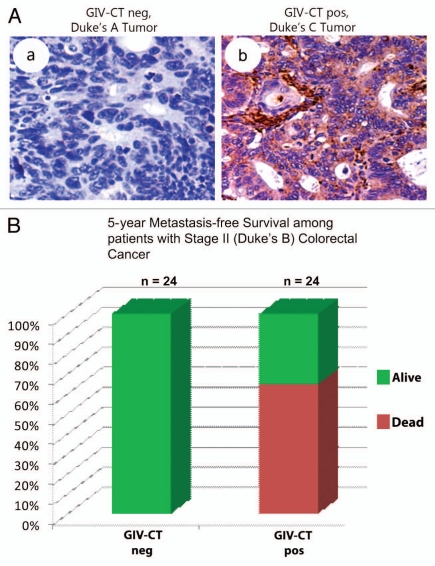
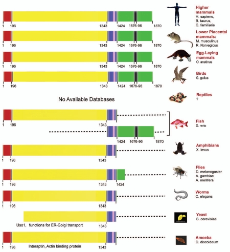
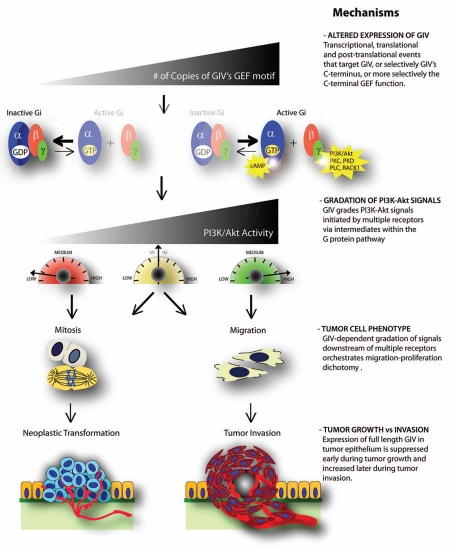
GIV/Girdin is a multidomain signaling molecule that enhances PI3K-Akt signals downstream of both G protein-coupled and growth factor receptors. We previously reported that GIV triggers cell migration via its C-terminal guanine-nucleotide exchange factor (GEF) motif that activates Gαi. Recently we discovered that GIV's C-terminus directly interacts with the epidermal growth factor receptor (EGFR), and when its GEF function is intact, a Gαi-GIV-EGFR signaling complex assembles. By coupling G proteins to growth factor receptors, GIV is uniquely poised to intercept the incoming receptor-initiated signals and modulate them via G protein intermediates. Subsequent work has revealed that expression of the highly specialized C-terminus of GIV undergoes a bipartite dysregulation during oncogenesis-full length GIV with an intact C-terminus is expressed at levels ~20-50-fold above normal in highly invasive cancer cells and metastatic tumors, but its C-terminus is truncated by alternative splicing in poorly invasive cancer cells and non-invasive tumors. The consequences of such dysregulation on graded signal transduction and cellular phenotypes in the normal epithelium and its implication during tumor progression are discussed herein. Based on the fact that GIV grades incoming signals initiated by ligand-activated receptors by linking them to cyclical activation of G proteins, we propose that GIV is a molecular rheostat for signal transduction.


Figures

Figure 1
Figure 1
GIV is a multidomain signaling molecule with key interacting partners. (A) Functional domains of GIV. The N-terminal Hook domain (red) interacts with microtubules, the coiled-coil domain (yellow) mediates homodimerization, the Gα-binding domain (GBD, blue) interacts with α-subunits of Gαi, a lipid (PI4P)-binding domain (pink) mediates interaction with the Golgi and the plasma membranes,, and the extreme C-terminus (green) interacts with EGFR, Akt kinase and actin., The key regulatory component of GIV, i.e., the GEF domain (black) with which GIV interacts and activates Gαi, is located within the C-terminus. (B) GIV interacts with multiple signaling molecules and cytoskeletal proteins. GIV/Girdin triggers migration and enhances PI3K-Akt signals downstream of a variety of growth factor receptors (VEGFR, EGFR,, IGFR and InsR7,41). GIV is also required for enhancement of PI3K-Akt signals downstream of GPCRs.,, GIV's C-terminal GEF domain activates Gαi and couples G proteins to multiple ligand-activated receptors. GIV is both an enhancer of Akt kinase and a substrate of the kinase, perhaps providing a feedback loop within the ‘receptor-GIV-PI3K-Akt’ signaling axis. GIV also interacts with 3 key structural components—actin, microtubules (MT) and PI4P-enriched membranes (Golgi and the PM) which serve to further compartmentalize and target GIV-dependent signaling pathways. (C) The Gαi-GIV interface is a unique therapeutic target. GIV is the first non-receptor GEF for heterotrimeric G proteins that works via a novel, evolutionarily conserved motif. The structure of GIV's GEF motif in complex with Gαi3 was initially modeled based on its homology with the synthetic KB-752 peptide in complex with Gαi1 and subsequently validated by site directed mutagenesis. GIV's GEF motif (red) docks on the hydrophobic cleft formed between Gαi3's α3 helix and the switch II (gold surface) via F1685. In addition, W258 in the α3/β5 loop of Gαi3 is an essential structural determinant that renders the G protein sensitive to activation by GIV. Importantly, the conservative substitution of W258 for F disrupts Gαi's ability to bind GIV, but it does not perturb its interaction with other binding partner such as Gβγ, GDIs, GAPs or GPCRs, which demonstrates that the Gαi-GIV interaction can be selectively targeted without compromising other functions of Gαi subunits. The uniqueness of the Gαi-GIV molecular interface, the selectivity of the structural determinants required to assemble it and its critical role in promoting the prometastatic functions associated with GIV expression in tumor cells make it a novel and attractive pharmacological target in cancer therapeutics.
Figure 2
Figure 2
GIV links G protein to ligand activated EGFR and regulates EGFR localization, EGFR degradation and EGF signaling. Schematic illustration of the EGFR itinerary and signaling profiles in cells expressing wild-type GIV with an intact GEF motif (left) and those expressing a GEF-deficient mutant of GIV (right). When GIV's GEF motif is intact, activation of Gαi is coupled to the ligand-activated EGFR via GIV, the duration and extent of receptor autophosphorylation and receptor association with the PM is enhanced, and the rate of receptor internalization is delayed. However, once internalized, the receptor rapidly transits through the endolysosomal compartments and receptor degradation is accelerated in lysosomes. By contrast, when GIV's GEF motif is disabled, Gαi is not activated in the vicinity of the receptor, receptor autophosphorylation is reduced, and it is rapidly internalized within endosomes where it stays for longer duration due to delayed receptor degradation. As a direct consequence of differential receptor association with the PM-actin bed, PM-based motogenic (PI3K-Akt and PLCγ1) signals are enhanced only when GIV's GEF motif is intact, whereas when GIV's GEF domain is disabled, mitogenic (c-Src, STAT5b and ERK1/2) signals are sustained from intracellular membranes.
Figure 3
Figure 3
Expression of GIV is dysregulated during cancer progression. Schematic illustration of changes in GIV expression in epithelial cells during transition from normal to cancer and during metastatic progression is displayed. While a non-invasive tumor with poor metastatic potential is largely comprised of poorly invasive tumor cells that express GIVΔCT, invasive tumors with high metastatic potential are populated by highly invasive tumor cells that overexpress full-length GIV.
Figure 4
Figure 4
Expression of full-length GIV increases correlation with metastatic progression and prognosticates metastasis-free survival in cancer patients. (A) Cells expressing a C-terminally truncated GIV (GIV-CT neg) dominate non-invasive tumors (a), whereas those with increased fulllength GIV (GIV-CT pos) are found in invasive tumors (b). Paraffin embedded human colon cancer samples were analyzed for full-length GIV by immunohistochemistry using GIV-CT Ab. Upper parts display representative fields from a non-invasive (Duke's A) tumor of early clinical stage (a) and invasive (Duke's C) tumors of late clinical stages (b). Non-invasive Duke's A tumor cells (a) stain negatively for GIV CT whereas invasive Duke's C tumors (b) are strongly positive. The percent of GIV-CT-positive tumors increases with increasing clinical stage of colorectal carcinoma-0% for Dukes A (early-staged tumor localized to mucosa), ∼48% for B (intermediate-staged tumor limited to muscularis propria) and 100% for C (advanced tumor spreads to local lymph nodes) and D (tumors with distant metastases). Parts reproduced with permission from Ghosh et al. MBoC, 2010. (B) Expression of full-length GIV prognosticates 5-year metastasis-free survival among patients with Stage II (Intermediate, Duke's B) colorectal carcinoma. Paraffin embedded human colon cancer samples of Duke's clinical stage B2 were analyzed for full-length GIV as in (A). Staining was scored as negative or positive by three independent observers blinded to patient outcome and stage with >95% congruency. At the 5-year mark, survival was 100% in the GIV-CT-negative group and 62 ± 9% (mean ± SE, p = 6 × 10⁻⁵) in the GIV-CT-positive group. Unpaired t-test revealed that the GIV-fl-negative and GIV-fl-positive subgroups were otherwise similar with respect to mean age at diagnosis and gender ratio.
Figure 5
Figure 5
The C-terminus of GIV which contains the EGFR/Akt/Actin-binding and GEF domains is evolutionarily young and evolved independently from the N-terminus. The various domains of mammalian GIV are color-coded as in Figure 1A. The various domains present in GIV orthologs in different species are displayed. The N-terminus of GIV (comprised of the hook, coiled-coil and G-binding domains) evolved early and is present in worms and flies. Although unicellular organisms (yeast and amoeba) do not have a clear ortholog of GIV, two proteins (Uso1 and interaptin) share some degree of similarity. The C-terminus of GIV evolved as part of a separate protein for the first time in fish. The N- and C-termini fused together as one GIV molecule in birds. Thus, birds and mammals express full-length GIV with the highly specialized C-terminus.
Figure 6
Figure 6
Working model for how GIV serves as a molecular rheostat during tumor progression. Schematic representation (from top to bottom) of how alterations in GIV expression balances mitosis and migration during tumor progression via intermediate steps of altered activation of G proteins and modulation of signals initiated by growth factor receptors. The molecular mechanism(s) that govern each step are listed on the right. Multiple mechanisms at transcriptional, translational and post-translational levels contribute to changes in the expression of GIV, or selectively it's C-terminus in the tumor epithelium, thereby directly affecting the number (#) of functionally intact copies of GIV's GEF motif (top). As a direct consequence of this, the rate-limiting step in cyclical activation of G proteins is proportionately altered over a broad range, in-continuum, from slow-cycling state in which the G protein spends longer duration in the GDP-bound inactive state to rapid-cycling state in which the G protein spends longer duration in the GTP-bound active state. This translates into graded enhancement of PI3K activity via G protein intermediates (Gβγ) and modulation of a variety of other motogenic and mitogenic signals by coupling of G protein activation to ligand-activated EGFR. Presence or absence of GIV's GEF function results in amplification of different sets of signaling programs and thereby, triggers migration-proliferation dichotomy—In the presence of an intact GEF motif, G proteins are activated, motogenic signals are enhanced and cell migration is triggered, whereas in the absence of a functional GEF motif, G proteins remain inactive, mitogenic signals are enhanced and mitosis is triggered. Expression of full-length GIV, and thereby the number of copies of functional GEF motifs is decreased early during tumor growth and increased later during tumor invasion, influencing both tumor size and metastasis.

References

    1. Le-Niculescu H, Niesman I, Fischer T, DeVries L, Farquhar MG. Identification and characterization of GIV, a novel Galpha i/s-interacting protein found on COPI, endoplasmic reticulum-Golgi transport vesicles. J Biol Chem. 2005;280:22012–22020. - PubMed
    1. Simpson F, Martin S, Evans TM, Kerr M, James DE, Parton RG, et al. A novel hook-related protein family and the characterization of hook-related protein 1. Traffic. 2005;6:442–458. - PubMed
    1. Anai M, Shojima N, Katagiri H, Ogihara T, Sakoda H, Onishi Y, et al. A novel protein kinase B (PKB)/AKT-binding protein enhances PKB kinase activity and regulates DNA synthesis. J Biol Chem. 2005;280:18525–18535. - PubMed
    1. Enomoto A, Murakami H, Asai N, Morone N, Watanabe T, Kawai K, et al. Akt/PKB regulates actin organization and cell motility via Girdin/APE. Dev Cell. 2005;9:389–402. - PubMed
    1. Enomoto A, Ping J, Takahashi M. Girdin, a novel actin-binding protein, and its family of proteins possess versatile functions in the Akt and Wnt signaling pathways. Ann NY Acad Sci. 2006;1086:169–184. - PubMed

Publication types

MeSH terms

Substances