. 2010 Mar 26;2(4):796-825.
doi: 10.3390/v2040796.

Alphavirus Entry and Membrane Fusion

Affiliations

Alphavirus Entry and Membrane Fusion

Margaret Kielian et al. Viruses. .

Abstract

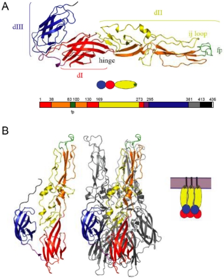
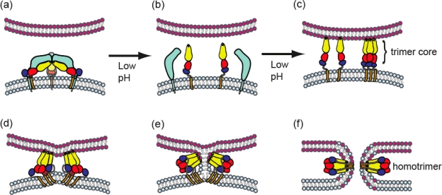
The study of enveloped animal viruses has greatly advanced our understanding of the general properties of membrane fusion and of the specific pathways that viruses use to infect the host cell. The membrane fusion proteins of the alphaviruses and flaviviruses have many similarities in structure and function. As reviewed here, alphaviruses use receptor-mediated endocytic uptake and low pH-triggered membrane fusion to deliver their RNA genomes into the cytoplasm. Recent advances in understanding the biochemistry and structure of the alphavirus membrane fusion protein provide a clearer picture of this fusion reaction, including the protein's conformational changes during fusion and the identification of key domains. These insights into the alphavirus fusion mechanism suggest new areas for experimental investigation and potential inhibitor strategies for anti-viral therapy.


Figures

Figure 1.
Figure 1.
The alphavirus membrane fusion protein E1 in the pre- and post-fusion conformations. (a) The prefusion structure of the SFV E1* ectodomain. The three domains of E1 are shown, with DI in red, the two insertions (into DI) that comprise DII shown in yellow and orange, and DIII in blue. The fusion peptide loop (fp) at the tip of domain II is in green, the DI-DIII linker is in purple, and the positions of the ij loop and hinge are indicated. Below the structure is a cartoon view of E1* with DI, II and III colored in red, yellow and blue, respectively and the fusion loop shown as a green star. The bottom part of the panel shows a linear diagram of the E1 sequence colored to match the structure above and labeled to indicate the domain boundaries. The stem is shown in grey, the TM domain in black, and the cytoplasmic tail of E1 in white. [PDB 2ALA, references 10,12] (b) The post-fusion structure of the E1 ectodomain. One E1* subunit from the homotrimer is shown in the left panel, colored as in A. The E1* homotrimer is shown in the middle panel with one E1* subunit colored as in A and the other two E1* subunits shown in light grey. DIII (blue) and the stem (dark grey) extend along the core trimer towards the fusion loops (green). The cartoon in the right panel illustrates the hairpin conformation and depicts the full-length membrane-inserted E1 homotrimer with the complete stem (grey), TM domain (black) and fused membrane (light purple). [PDB 1RER, reference 13] Figure reprinted from Virology, 344, Kielian, M., Class II virus membrane fusion proteins, p. 40, Copyright (2006), with permission from Elsevier.
Figure 2.
Figure 2.
Model for stages of the alphavirus membrane fusion reaction. (a) Virus particle in the pre-fusion state. The virus membrane, depicted in light blue, contains a trimer of E2-E1 heterodimers, with E2 in light blue and E1 colored as in Figure 1. The target membrane is shown in pink. The fusion protein E1 is in a metastable conformation. (b) Triggering. Upon exposure to low pH, dissociation of the E2-E1 heterodimer occurs, exposing the E1 fusion loop. The disposition of E2 after heterodimer dissociation is unknown. (c) The fusion loop inserts in the target membrane through a low pH and cholesterol-dependent mechanism. A core trimer is formed by DI and DII. (d-e). In a pH-independent interaction, DIII and the stem region are folded against the core trimer in the groove formed by two E1 proteins. The distortion of the target membrane by fusion loop insertion, the fold-back of DIII and stem, and the cooperative action of several trimers (of which only two are shown) are proposed to provide the force to mediate membrane fusion. (e) Fusion proceeds through a hemifusion step in which the two outer leaflets merge. (f) E1 forms the final stable post-fusion homotrimer, in which the fusion loops and the transmembrane domains are located at the same side of the molecule. Concomitantly, this refolding drives complete fusion via formation of the fusion pore. Figure reprinted from Trends in Microbiology, 17, Sanchez-San Martin, C., Liu, C. Y., and Kielian, M., Dealing with low pH: entry and exit of alphaviruses and flaviviruses, p. 517, Copyright (2009), with permission from Elsevier.

Similar articles

Cited by

References

    1. Marsh M, Helenius A. Virus entry: open sesame. Cell. 2006;124:729–740. - PMC - PubMed
    1. Gruenberg J. Viruses and endosome membrane dynamics. Curr Opin Cell Biol. 2009;21:582–588. - PubMed
    1. Sieczkarski SB, Whittaker GR. Viral entry. Curr Topics Microbiol Immunol. 2005;285:1–23. - PubMed
    1. Harrison SC. Viral membrane fusion. Nat Struct Mol Biol. 2008;15:690–698. - PMC - PubMed
    1. White JM, Delos SE, Brecher M, Schornberg K. Structures and mechanisms of viral membrane fusion proteins: multiple variations on a common theme. Crit Rev Biochem Mol Biol. 2008;43:189–219. - PMC - PubMed

LinkOut - more resources