

Fetomaternal microchimerism: Some answers and many new questions
- PMID: 21547031
- PMCID: PMC3084951
- DOI: 10.4161/chim.2.1.14692
Fetomaternal microchimerism: Some answers and many new questions
Abstract
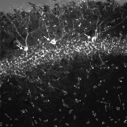
The transfer of fetal cells into mothers during pregnancy and their organ specific integration is a well recognized phenomenon in placental vertebrates. Recently, it has been reported that some fetal cells found in the mothers have progenitor cell-like features such as multilineage differentiation potential and as a consequence they were termed pregnancy associated progenitor cells (PAPC). The multilineage differentiation potential suggested some level of cellular plasticity, which these cells share with other stem or progenitor cells. In this context, we have shown that PAPCs indeed express neural stem cell and markers for developing neurons in the brain and that PAPCs morphologically mature into neurons over time. The stem/progenitor properties of PAPCs raises the hope that they might be valuable for studying the functional integration of foreign cells into preexisting tissues and organs, for example in cellular therapies. The functional integration of transplanted cells and their connectivity to the host circuitry is still a major bottleneck in cellular therapies particularly for the brain. The animal models of fetomaternal microchimerism might provide valuable insights into the mechanism how cells survive, migrate, integrate and differentiate in a foreign environment of a host. This review discusses some of the recent findings in the field of fetomaternal microchimerism. It also tries to identify some major gaps of knowledge and raises some questions resulting from the recent advances. Studying fetomaternal microchimerism and the properties of PAPCs in greater detail might pave the way to advance cell based regenerative medicine as well as transplantation medicine.
Figures
Comment on
-
Pregnancy-associated progenitor cells differentiate and mature into neurons in the maternal brain.Stem Cells Dev. 2010 Dec;19(12):1819-30. doi: 10.1089/scd.2010.0046. Epub 2010 Sep 13. Stem Cells Dev. 2010. PMID: 20707697
References
-
- Greger WP, Steele MR. Human fetomaternal passage of erythrocytes. N Engl J Med. 1957;256:158–161. - PubMed
-
- Desai RG, Creger WP. Maternofetal passage of leukocytes and platelets in man. Blood. 1963;21:665–673. - PubMed
-
- Lee RE, Vazquez JJ. Immunocytochemical evidence for transplacental passage of erythrocytes. Lab Invest. 1962;11:580–584. - PubMed
-
- Tuffrey M, Bishun NP, Barnes RD. Porosity of the mouse placenta to maternal cells. Nature. 1969;221:1029–1030. - PubMed
LinkOut - more resources
Full Text Sources