. 2011 Apr 25;6(4):e18984.
doi: 10.1371/journal.pone.0018984.

Dispersal of Group A streptococcal biofilms by the cysteine protease SpeB leads to increased disease severity in a murine model

Affiliations

Dispersal of Group A streptococcal biofilms by the cysteine protease SpeB leads to increased disease severity in a murine model

Kristie L Connolly et al. PLoS One. .

Abstract

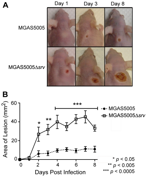
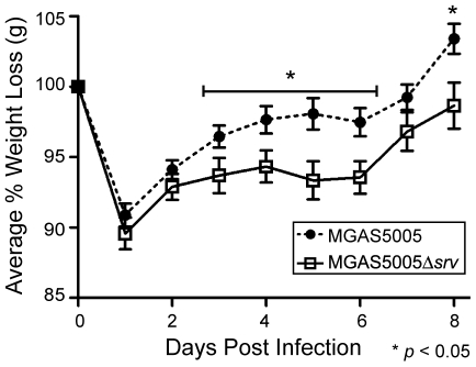
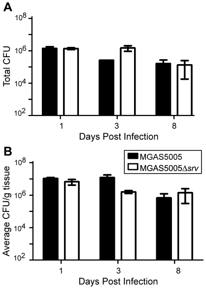
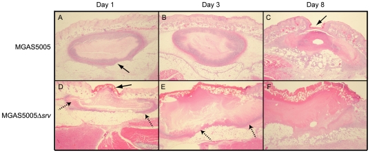
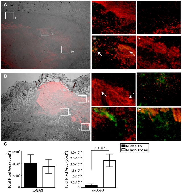
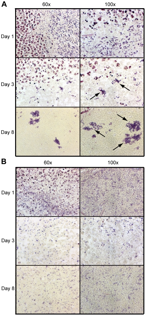
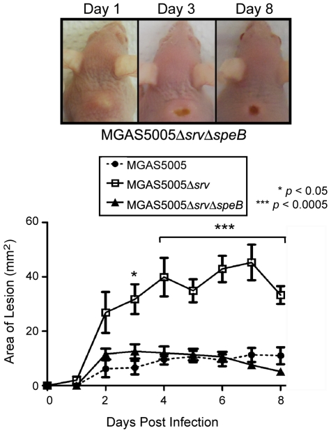
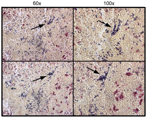
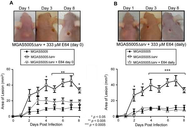
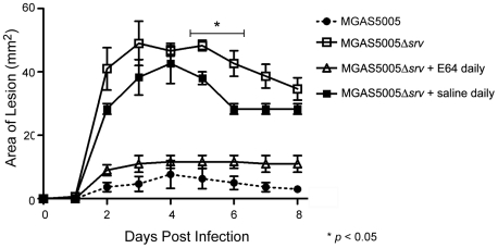
Group A Streptococcus (GAS) is a Gram-positive human pathogen best known for causing pharyngeal and mild skin infections. However, in the 1980's there was an increase in severe GAS infections including cellulitis and deeper tissue infections like necrotizing fasciitis. Particularly striking about this elevation in the incidence of severe disease was that those most often affected were previously healthy individuals. Several groups have shown that changes in gene content or regulation, as with proteases, may contribute to severe disease; yet strains harboring these proteases continue to cause mild disease as well. We and others have shown that group A streptococci (MGAS5005) reside within biofilms both in vitro and in vivo. That is to say that the organism colonizes a host surface and forms a 3-dimensional community encased in a protective matrix of extracellular protein, DNA and polysaccharide(s). However, the mechanism of assembly or dispersal of these structures is unclear, as is the relationship of these structures to disease outcome. Recently we reported that allelic replacement of the streptococcal regulator srv resulted in constitutive production of the streptococcal cysteine protease SpeB. We further showed that the constitutive production of SpeB significantly decreased MGAS5005Δsrv biofilm formation in vitro. Here we show that mice infected with MGAS5005Δsrv had significantly larger lesion development than wild-type infected animals. Histopathology, Gram-staining and immunofluorescence link the increased lesion development with lack of disease containment, lack of biofilm formation, and readily detectable levels of SpeB in the tissue. Treatment of MGAS5005Δsrv infected lesions with a chemical inhibitor of SpeB significantly reduced lesion formation and disease spread to wild-type levels. Furthermore, inactivation of speB in the MGAS5005Δsrv background reduced lesion formation to wild-type levels. Taken together, these data suggest a mechanism by which GAS disease may transition from mild to severe through the Srv mediated dispersal of GAS biofilms.


Conflict of interest statement

Competing Interests: The authors have declared that no competing interests exist.

Figures

Figure 1
Figure 1. Allelic replacement of srv lead to increased lesion size in a murine subcutaneous infection model.
(A) Groups of 10 mice (Crl:SKH1-hrBR) were challenged subcutaneously with ∼2.0×10⁸ CFU (0.1 ml) of either MGAS5005 or MGAS5005Δsrv. Representative images of lesions formed at 1, 3 and 8 dpi are shown. (B) The area of the lesion formed (mm2) was measured with a caliper daily. Lesions formed by MGAS5005Δsrv were significantly larger (p≤0.05) than those formed by MGAS5005 by 2 dpi (Student's t-test).
Figure 2
Figure 2. Average percentage of mouse weight loss following GAS infection.
Groups of 10 mice were challenged subcutaneously with ∼2.0×10⁸ CFU (0.1 ml) of either MGAS5005 or MGAS5005Δsrv. The percentage of weight lost was monitored for 8 dpi. Mice infected with MGAS5005Δsrv weighed significantly less on 5/8 dpi (*p≤0.05).
Figure 3
Figure 3. Bacterial load recovered from excised lesions.
Lesions from mice infected with either MGAS5005 or MGAS5005Δsrv (n = 3 mice/strain) were excised at 1, 3 and 8 dpi, weighed and homogenized for replicate plating. No significant difference in (A) total CFU recovered or (B) CFU/g was observed at 1, 3, and 8 dpi.
Figure 4
Figure 4. Histopathology of excised lesions from MGAS5005 and MGAS5005Δsrv infections.
Lesions were surgically excised at days 1, 3, and 8 post infection. 10 µm sections were subjected to H&E staining. Representative low-magnification images (2×) from each time point are shown. (A,B) Infection with MGAS5005 resulted in the formation of a subcutaneous abscess (arrow) that was well delineated by fibrin (pink border) and PMNs (purple border). (C) By 8 dpi, the abscess had ruptured and formed a cutaneous lesion that showed signs of healing (arrow). (D) MGAS5005Δsrv infection resulted in a cutaneous lesion (arrow). (D & E) Note the subcutaneous abscess was less contained by colocalized fibrin and PMNs (dashed arrows). (E) The cutaneous lesion grew in size and did not show any appreciable healing by 8 dpi (F).
Figure 5
Figure 5. Immunofluorescent antibody staining revealed detectable levels of SpeB throughout MGAS5005Δsrv infected tissue as compared to MGAS5005 infected tissue.
Subcutaneous abscesses from (A) MGAS5005 and (B) MGAS5005Δsrv infections were excised 1 dpi, sectioned, and stained with rabbit anti-SpeB sera and goat anti-GAS sera, and the appropriate fluorescent secondary antibody conjugate. (A, B) DIC/fluorescent images (4×) from an MGAS5005 infected animal (A) and an MGAS5005Δsrv infected animal (B) show the distribution of GAS (red) throughout the abscess. Randomly selected areas throughout the abscesses were examined for the colocalization of GAS and SpeB (20×, i–iv). MGAS5005 was readily detected (Ai–iv), but SpeB (green) was rarely detected in MGAS5005 infected samples (arrows, Aiii). In contrast, SpeB was detected in the presence of MGAS5005Δsrv throughout the infected samples (Bi–iv). Colocalized SpeB and GAS appear yellow. Representative images are shown. (C) Average total area of pixels (pixels2) was calculated for anti-GAS and anti-SpeB staining in the representative images shown of MGAS5005 and MGAS5005Δsrv. Comparable amounts of anti-GAS staining was observed, however, there is significantly more anti-SpeB staining in MGAS5005Δsrv images compared to MGAS5005 (* p<0.01).
Figure 6
Figure 6. Gram-staining of lesion tissue sections revealed the presence of MGAS5005 microcolonies (biofilms).
10 µm sections of lesion tissue collected 1, 3, and 8 dpi were subjected to Gram-staining. (A) MGAS5005 infected samples contained microcolonies of adherent GAS which were visible by 3 dpi (arrows). These microcolonies are reminiscent of biofilms and appeared to increase in size by 8 dpi. (B) MGAS5005Δsrv infected samples contained randomly dispersed GAS throughout the field of view. Microcolonies were largely absent. The same view of single day images are shown at 60× and 100× magnification. Representative images are shown.
Figure 7
Figure 7. Allelic replacement of speB in the MGAS5005Δsrv background resulted in significantly decreased lesion size.
Representative images of lesions formed in mice at 1, 3 and 8 days following subcutaneous infection with ∼2×10⁸ CFU (0.1 ml) of MGAS5005ΔsrvΔspeB. Lesion development (mm2) was monitored over 8 days using a caliper (n = 10 mice/strain). A significant reduction in lesion size was observed in MGAS5005ΔsrvΔspeB infected mice (p<0.05). The size of lesions observed in MGAS5005 infected mice vs. MGAS5005ΔsrvΔspeB was not significantly different.
Figure 8
Figure 8. Microcolony formation is observed in MGAS5005ΔsrvΔspeB infected tissue.
Representative images of Gram-stained sections (10 µm thick) collected from two MGAS5005ΔsrvΔspeB infected mice at 8 dpi. MGAS5005ΔsrvΔspeB microcolonies (arrows) were present in the edema at the site of infection.
Figure 9
Figure 9. Use of the chemical inhibitor of cysteine proteases E64 significantly reduced lesion size in MGAS5005Δsrv infected animals.
Representative images of lesions formed in mice at 1, 3 and 8 days following subcutaneous infection with ∼2×10⁸ CFU of MGAS5005Δsrv. (A) The infecting dose of MGAS5005Δsrv was suspended in 333 uM E64 (0.1 ml), and lesion development (mm2) was monitored over 8 days (n = 10 mice). A significant reduction in lesion formation was observed when E64 was inoculated with the infecting dose of MGAS5005Δsrv compared to inoculation with MGAS5005Δsrv alone (p<0.05). (B) Following inoculation of animals with E64+MGAS5005Δsrv as before, an additional inoculation of 333 µM E64 (0.1 mL) was injected directly into the abscess each day following infection (n = 10). A significant reduction in lesion size was observed with E64 treated animals forming lesions roughly equivalent in size to untreated MGAS5005 infected animals.
Figure 10
Figure 10. Daily wound irrigation not responsible for the reduction in lesion size observed in E64 treated animals.
Lesion size in saline treated animals (n = 3) was significantly reduced at 5 and 6 dpi compared to MGAS5005Δsrv infected animals (p<0.05), however, lesions in saline treated animals were statistically larger than those in E64 treated animals at 2–8 dpi (p<0.05).

Similar articles

Cited by

References

    1. Gabillot-Carre M, Roujeau JC. Acute bacterial skin infections and cellulitis. Curr Opin Infect Dis. 2007;20:118–123. - PubMed
    1. Martin JM, Green M. Group A streptococcus. Semin Pediatr Infect Dis. 2006;17:140–148. - PubMed
    1. Rogers RL, Perkins J. Skin and soft tissue infections. Prim Care. 2006;33:697–710. - PubMed
    1. Stevens DL, Bisno AL, Chambers HF, Everett ED, Dellinger P, et al. Practice guidelines for the diagnosis and management of skin and soft-tissue infections. Clin Infect Dis. 2005;41:1373–1406. - PubMed
    1. Cunningham MW. Pathogenesis of group A streptococcal infections. Clin Microbiol Rev. 2000;13:470–511. - PMC - PubMed

Publication types

MeSH terms