

Review
doi: 10.3748/wjg.v17.i16.2080.
Endoscopic ultrasonography findings in autoimmune pancreatitis
Affiliations
- PMID: 21547126
- PMCID: PMC3084392
- DOI: 10.3748/wjg.v17.i16.2080
Item in Clipboard
Review
Endoscopic ultrasonography findings in autoimmune pancreatitis
World J Gastroenterol.
.
Abstract
Endoscopic ultrasonography is an established diagnostic tool for pancreatic masses and chronic pancreatitis. In recent years there has been a growing interest in the worldwide medical community in autoimmune pancreatitis (AIP), a form of chronic pancreatitis caused by an autoimmune process. This paper reviews the current available literature about the endoscopic ultrasonographic findings of AIP and the role of this imaging technique in the management of this protean disease.
Keywords: Autoimmune; Endoscopic ultrasound; IgG4 cholangitis; Pancreatitis.
Figures
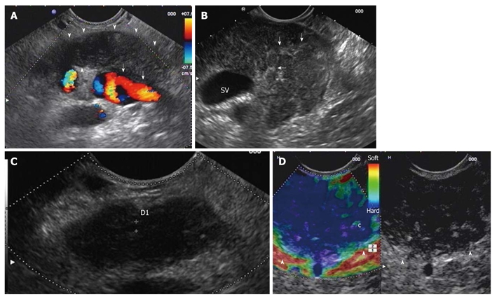
Diffuse form of autoimmune pancreatitis. A: Endoscopic ultrasonography (EUS) linear scanning shows a diffuse pancreatic enlargement (arrowheads) with echopoor echotexture, and with loss of interface with splenic vein (arrows); B: Parenchymal lobularity and hyperechoic strands (arrows) are visible in the enlarged gland; C: Pancreatic duct calliper is 1.8 mm; D: EUS-elastography demonstrates the diffuse pancreatic stiffness (arrowheads).
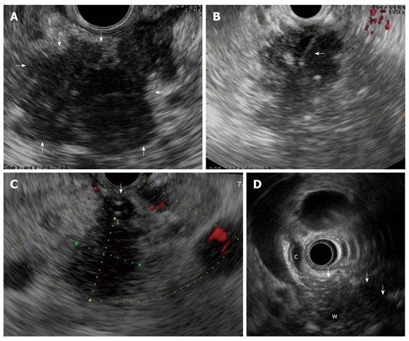
Focal form of autoimmune pancreatitis. A: Endoscopic ultrasonography (EUS) shows a focal lesion (arrows) of pancreatic head which is echopoor with hyperechoic strands; B: A EUS-guided fine needle aspiration is performed (arrow) for tissue characterization; C: Another case of focal autoimmune pancreatitis (AIP) with echopoor lesion of pancreatic head (between callipers) and marked echopoor thickening of the choledochal wall (arrow); D: In this case of focal AIP EUS shows a echopoor lesion (arrows) of pancreatic head, with upstream dilatation of both common bile duct (c) and pancreatic duct (w); notice the thickened choledochal wall.
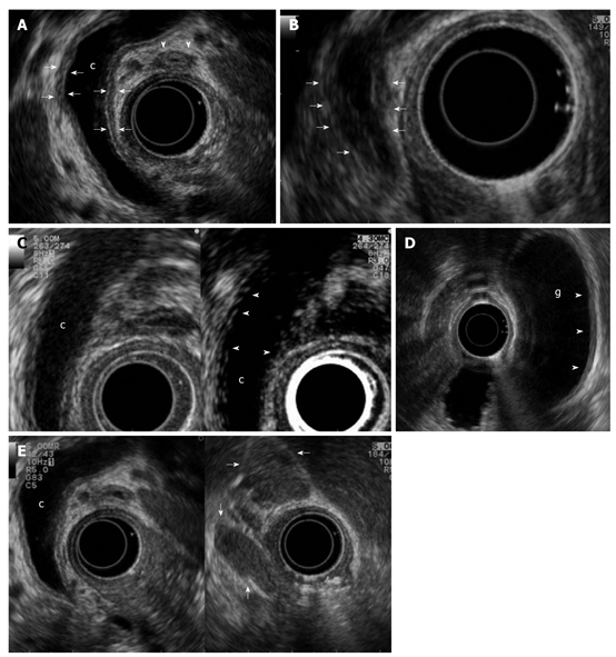
Biliary and peripancreatic findings in autoimmune pancreatitis. Autoimmune pancreatitis presenting with jaundice: A: Endoscopic ultrasonography (EUS) shows a dilated common bile duct (c) upstream to a distal funnel-shaped stenosis; EUS demonstrates the diffuse thickening of the biliary wall (between arrows) with “sandwich-pattern”, either of common bile duct or of cystic duct (arrowheads). This thickening is equally visible both in the dilated region of the common bile duct; B: In the distal strictured tract (arrows); C: After contrast administration (Sonovue, Bracco) the biliary wall shows an early and persistent enhancement (arrowheads); D: EUS shows the same thickening of the gallbladder (g) wall (arrowheads); E: Enlarged lymph nodes to the hepatic hylum (arrows).
References
-
- Chang DK, Nguyen NQ, Merrett ND, Dixson H, Leong RW, Biankin AV. Role of endoscopic ultrasound in pancreatic cancer. Expert Rev Gastroenterol Hepatol. 2009;3:293–303. - PubMed
-
- Kahl S, Glasbrenner B, Leodolter A, Pross M, Schulz HU, Malfertheiner P. EUS in the diagnosis of early chronic pancreatitis: a prospective follow-up study. Gastrointest Endosc. 2002;55:507–511. - PubMed
-
- Buscarini E, Frulloni L, De Lisi S, Falconi M, Testoni PA, Zambelli A. Autoimmune pancreatitis: a challenging diagnostic puzzle for clinicians. Dig Liver Dis. 2010;42:92–98. - PubMed
-
- Japan Pancreas Society. Diagnostic criteria for autoimmune pancreatitis. J Jpn Pancreas Soc. 2002;17:585–587.
Publication types
MeSH terms
LinkOut - more resources
Full Text Sources
Medical
Miscellaneous
