

PET Imaging of Integrin αVβ3 Expression
- PMID: 21547152
- PMCID: PMC3086612
- DOI: 10.7150/thno/v01p0048
PET Imaging of Integrin αVβ3 Expression
Abstract
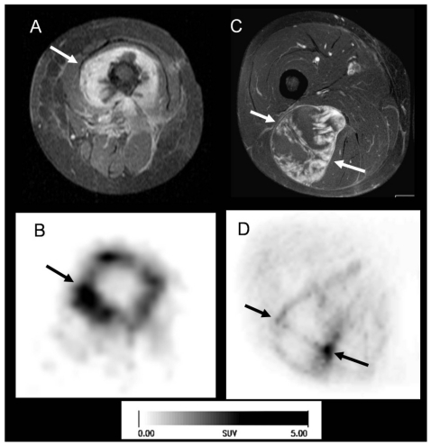
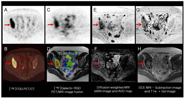
PET imaging of integrin αvβ3 expression has been studied intensely by the academia and recently also by the industry. Imaging of integrin αvβ3 expression is of great potential value, as the integrin αvβ3 is a key player in tumor metastasis and angiogenesis. Therefore PET imaging of this target might be a suitable in-vivo biomarker of angiogenesis and metastatic potential of tumors. In this manuscript, the various strategies for PET imaging of the integrin αvβ3 will be summarized, including monomeric and multimeric radiolabelled RGD peptides and nanoparticles. While most experiments have been performed using preclinical tumor models, more and more clinical results on PET imaging of αvβ3 expression are available and will be discussed in detail. However, while a multitude of radiotracer strategies have been successfully evaluated for PET imaging of αvβ3, the ultimate clinical value of this new imaging biomarker still has to be evaluated in large clinical trials.
Keywords: PET; angiogenesis; integrin αvβ3; metastasis; molecular imaging.
Conflict of interest statement
Conflict of Interest: The authors have declared that no conflict of interest exists.
Figures

References
-
- Folkman J. Angiogenesis in cancer, vascular, rheumatoid and other disease. Nat Med. 1995;1:27–31. - PubMed
-
- Risau W. Mechanisms of angiogenesis. Nature. 1997;386:671–4. - PubMed
-
- Kerbel RS. Antiangiogenic therapy: a universal chemosensitization strategy for cancer? Science. 2006;312:1171–5. - PubMed
-
- Hurwitz H, Fehrenbacher L, Novotny W, Cartwright T, Hainsworth J, Heim W. et al. Bevacizumab plus irinotecan, fluorouracil, and leucovorin for metastatic colorectal cancer. N Engl J Med. 2004;350:2335–42. - PubMed
-
- Galbraith SM. Antivascular cancer treatments: imaging biomarkers in pharmaceutical drug development. Br J Radiol. 2003;76:S83–6. - PubMed
LinkOut - more resources
Full Text Sources
Other Literature Sources
