Distinguishing benign from malignant parotid gland tumours: low-dose multi-phasic CT protocol with 5-minute delay
- PMID: 21547526
- PMCID: PMC3128264
- DOI: 10.1007/s00330-011-2101-y
Distinguishing benign from malignant parotid gland tumours: low-dose multi-phasic CT protocol with 5-minute delay
Abstract
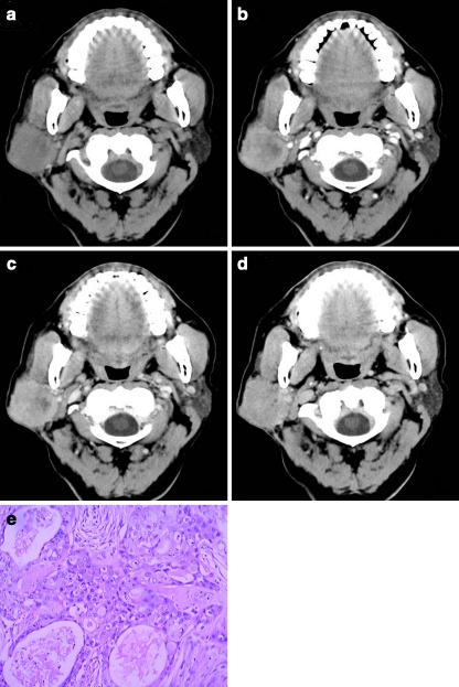
Objectives: To explore the percentage enhancement wash-out ratio (PEW) and relative PEW (RPEW) of low-dose multi-phasic computed tomography (CT) in distinguishing benign from malignant parotid gland tumours.
Methods: This study was approved by the ethics committee, and informed patient consent was obtained. 51 patients with parotid tumours proven by histopathology received CT, including 18 with pleomorphic adenomas, 14 with Warthin's tumours and 19 with malignant tumours. Size and attenuation of parotid tumours were measured. Compared with 5-min attenuation, the 30-s and 90-s PEW (PEW(30,) PEW(90)) and RPEW (RPEW(30), RPEW(90)) were calculated.
Results: There was a significant difference in PEW(30), RPEW(30), PEW(90) and RPEW(90) in the parotid neoplasms groups (P < 0.01), and statistical significance existed simultaneously in pleomorphic adenomas vs malignant tumours and Warthin's tumours vs malignant tumours according to SNK-q test. The optimal diagnosis results of malignancy with 100% specificity (32/32) was obtained by using a combination of the following criteria: -70% > PEW(30) < 36%, -30% > PEW(30) < 19%, PEW(90) > 12%, and the sensitivity (74%) for diagnosis of malignancy was yield.
Conclusions: Wash-out ratio may assist in differentiating the benign from malignant parotid gland tumours. Combining the percentage of enhanced wash-out ratios of CT protocols can yield diagnostic results for malignancy.
Figures

Similar articles
-
Diagnostic value of CT perfusion imaging for parotid neoplasms.Dentomaxillofac Radiol. 2014;43(1):20130237. doi: 10.1259/dmfr.20130237. Epub 2013 Nov 1. Dentomaxillofac Radiol. 2014. PMID: 24186883 Free PMC article.
-
Perfusion imaging of parotid gland tumours: usefulness of arterial spin labeling for differentiating Warthin's tumours.Eur Radiol. 2015 Nov;25(11):3247-54. doi: 10.1007/s00330-015-3755-7. Epub 2015 Apr 29. Eur Radiol. 2015. PMID: 25921591
-
Parotid gland tumors: comparison of conventional and diffusion-weighted MRI findings with histopathological results.Dentomaxillofac Radiol. 2021 May 1;50(4):20200391. doi: 10.1259/dmfr.20200391. Epub 2020 Dec 11. Dentomaxillofac Radiol. 2021. PMID: 33237812 Free PMC article.
-
Gray scale and Doppler ultrasonography of the benign tumors of parotid gland (pleomorphic adenoma and Warthin's tumor). Pictorial essay.Med Ultrason. 2010 Sep;12(3):238-44. Med Ultrason. 2010. PMID: 21203603 Review.
-
Contrast-Enhanced Ultrasound in the Differentiation between the Most Common Benign Parotid Gland Tumors: A Systematic Review and Meta-Analysis.J Clin Med. 2022 Dec 12;11(24):7360. doi: 10.3390/jcm11247360. J Clin Med. 2022. PMID: 36555976 Free PMC article. Review.
Cited by
-
Diagnostic value of CT perfusion imaging for parotid neoplasms.Dentomaxillofac Radiol. 2014;43(1):20130237. doi: 10.1259/dmfr.20130237. Epub 2013 Nov 1. Dentomaxillofac Radiol. 2014. PMID: 24186883 Free PMC article.
-
Sebaceous Lymphadenoma of Parotid: Imaging, Cytological, and Histological Findings in Detail.Case Rep Pathol. 2018 Apr 18;2018:2915907. doi: 10.1155/2018/2915907. eCollection 2018. Case Rep Pathol. 2018. PMID: 29850339 Free PMC article.
-
Basal cell adenoma and myoepithelioma of the parotid gland: patterns of enhancement at two-phase CT in comparison with Warthin tumor.Diagn Interv Radiol. 2019 Jul;25(4):285-290. doi: 10.5152/dir.2019.18337. Diagn Interv Radiol. 2019. PMID: 31120425 Free PMC article.
-
Value of Diffusion-Weighted Imaging Combined with Susceptibility-Weighted Imaging in Differentiating Benign from Malignant Parotid Gland Lesions.Med Sci Monit. 2018 Jul 4;24:4610-4616. doi: 10.12659/MSM.911185. Med Sci Monit. 2018. PMID: 29972148 Free PMC article.
-
A Preliminary Study of CT Texture Analysis for Characterizing Epithelial Tumors of the Parotid Gland.Cancer Manag Res. 2020 Apr 21;12:2665-2674. doi: 10.2147/CMAR.S245344. eCollection 2020. Cancer Manag Res. 2020. PMID: 32368145 Free PMC article.
References
-
- Nagler RM, Laufer D. Tumors of the major and minor salivary glands: review of 25 years of experience. Anticancer Res. 1997;17:701–707. - PubMed
-
- McGahan JP, Walter JP, Bernstein L. Evaluation of the parotid gland. Comparison of sialography, non-contrast computed tomography, and CT sialography. Radiology. 1984;52:453–458. - PubMed
-
- Choi DS, Na DG, Byun HS, et al. Salivary gland tumors: evaluation with two-phase helical CT. Radiology. 2000;214:231–236. - PubMed
MeSH terms
Substances
LinkOut - more resources
Full Text Sources
Medical