. 2011 Jun 6;8(3):940-8.
doi: 10.1021/mp2000583. Epub 2011 May 16.

Effects of noncovalent platinum drug-protein interactions on drug efficacy: use of fluorescent conjugates as probes for drug metabolism

Affiliations

Effects of noncovalent platinum drug-protein interactions on drug efficacy: use of fluorescent conjugates as probes for drug metabolism

Brad T Benedetti et al. Mol Pharm. .

Abstract

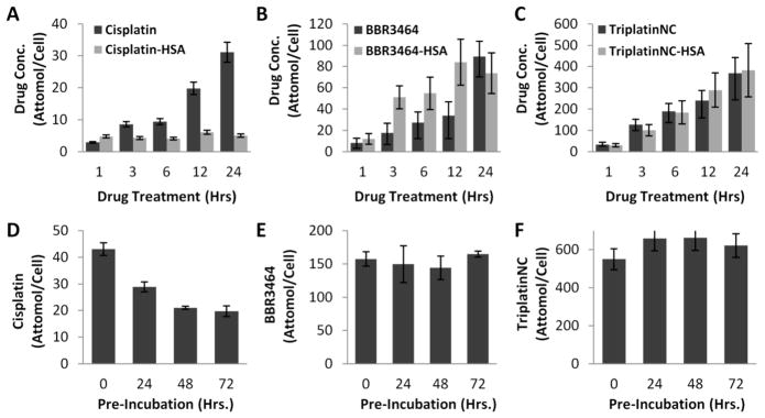
The overall efficacy of platinum based drugs is limited by metabolic deactivation through covalent drug-protein binding. In this study the factors affecting cytotoxicity in the presence of glutathione, human serum albumin (HSA) and whole serum binding with cisplatin, BBR3464, and TriplatinNC, a "noncovalent" derivative of BBR3464, were investigated. Upon treatment with buthionine sulfoximine (BSO), to reduce cellular glutathione levels, cisplatin and BBR3464-induced apoptosis was augmented whereas TriplatinNC-induced cytotoxicity was unaltered. Treatment of A2780 ovarian carcinoma cells with HSA-bound cisplatin (cisplatin/HSA) and cisplatin preincubated with whole serum showed dramatic decreases in cytotoxicity, cellular accumulation, and DNA adduct formation compared to treatment with cisplatin alone. Similar effects are seen with BBR3464. In contrast, TriplatinNC, the HSA-bound derivative (TriplatinNC/HSA), and TriplatinNC pretreated with whole serum retained identical cytotoxic profiles and equal levels of cellular accumulation at all time points. Confocal microscopy of both TriplatinNC-NBD, a fluorescent derivative of TriplatinNC, and TriplatinNC-NBD/HSA showed nuclear/nucleolar localization patterns, distinctly different from the lysosomal localization pattern seen with HSA. Cisplatin-NBD, a fluorescent derivative of cisplatin, was shown to accumulate in the nucleus and throughout the cytoplasm while the localization of cisplatin-NBD/HSA was limited to lysosomal regions of the cytoplasm. The results suggest that TriplatinNC can avoid high levels of metabolic deactivation currently seen with clinical platinum chemotherapeutics, and therefore retain a unique cytotoxic profile after cellular administration.


Figures

Figure 1
Figure 1
Chemical structures of cisplatin (top), BBR3464 (middle) and TriplatinNC (bottom).
Figure 2
Figure 2
(A) Effect of BSO on platinum drug-induced cytotoxicity in HCT116 cells. Subdiploid cell content was detected by PI-DNA staining. HCT116 cells were cultured with 10 μM cisplatin, 50 μM BBR3464, or 40 μM TriplatinNC for 48 h in the absence or presence BSO. Drugs were added to the medium after 1 h of treatment with BSO. (B) Effect of BSO on platinum drug cellular uptake in HCT116 carcinoma cell lines. HCT116 cells were cultured with 20 μM cisplatin for 16 h, 20 μM BBR3464 for 8 h, or 20 μM TriplatinNC for 3 h in the absence or presence of 500 μM BSO. Drugs were added to the medium after 1 h of treatment with BSO.
Figure 3
Figure 3
Influence of protein binding on platinum drug efficacy in A2780 ovarian carcinoma cells. (A) Cisplatin cytotoxicity (▲) in A2780 cells compared with the treatment of cisplatin/HSA (■). (B) BBR3464 cytotoxicity (▲) compared with the treatment of BBR3464/HSA (■). (C) TriplatinNC cytotoxicity (▲) compared with the treatment of TriplatinNC/HSA (■). (D–F) Platinum compounds were incubated with serum supplemented medium for 0–72 h. Cellular grown inhibition was then measured after treatment. Efficacy of cisplatin (D), BBR3464 (E), and TriplatinNC (F) following increased serum incubation times (0 (▲), 24 (■), 48 (◆), and 72 (●) h). A2780 cells were cultured in the indicated concentrations of each compound for 72 h. Percent growth inhibition was determined by MTT.
Figure 4
Figure 4
Influence of protein binding on platinum cellular uptake in A2780 ovarian carcinoma cells. A2780 cells were treated with IC90 concentrations of cisplatin and cisplatin/HSA (A), BBR3464 and BBR3464/HSA (B), TriplatinNC and TriplatinNC/HSA (C) for 0, 3, 6, 12, and 24 h. For serum incubation studies each platinum drug was preincubated with serum for (0–72 h). A2780 cells were then treated with IC90 concentrations of each compound ((D) cisplatin, (E) BBR3464, (F) TriplatinNC) for 24 h. Analysis of platinum content was performed on a Varian ICP-MS.
Figure 5
Figure 5
Influence of protein binding on DNA platination in A2780 ovarian carcinoma cells. A2780 cells were treated with IC90 concentrations of cisplatin and cisplatin/HSA (A), BBR3464 and BBR3464/HSA (B) for 24 h. For serum incubation studies each platinum drug was preincubated with serum for (0–72 h). A2780 cells were then treated with IC90 concentrations of each compound ((C) cisplatin, (D) BBR3464) for 24 h. Cellular DNA was extracted and analysis of platinum content was performed on a Varian ICP-MS.
Figure 6
Figure 6
Role of protein binding on the cellular localization and distribution of cisplatin and TriplatinNC. Confocal laser scanning micrographs (A, B) of A2780 cells. (A) (top row) Cisplatin-NBD (15 μM) (green) incubated with A2780 cells for 24 h and LysoTracker Red (75 nM) (red) for final 30 min of drug incubation. (A) (bottom row) Cisplatin-NBD/HSA (1 mg/mL) (green) incubated with A2780 cells for 24 h and LysoTracker Red (75 nM) (red) for final 30 min of drug incubation. Orange represents areas of drug–protein colocalization. (B) (top row) TriplatinNC-NBD (15 μM) (green) incubated with A2780 cells for 6 h and LysoTracker Red (75 nM) (red) for final 30 min of drug incubation. (B) (bottom row) TriplatinNC-NBD/HSA (1 mg/mL) (green) incubated with A2780 cells for 6 h and LysoTracker Red (75nM) (red) for final 30 min of drug incubation. Slides were mounted with Vectashield mounting medium containing DAPI (blue). Fluorescence was observed by confocal laser scanning microscopy (Zeiss LSM 510). (C) Chemical structures of TriplatinNC-NBD (top) and cisplatin-NBD (bottom).

References

    1. O’Dwyer P, Stevenson J, Johnson S. Clinical pharmacokinetics and administration of established platinum drugs. Drugs. 2000;59:19–27. - PubMed
    1. Decatris MP, Sundar S, O’Byrne KJ. Platinum-based chemotherapy in metastatic breast cancer: current status. Cancer Treat Rev. 2004;30:53–81. - PubMed
    1. Wong E, Giandomenico CM. Current status of platinum-based antitumor drugs. Chem Rev. 1999;99:2451–2466. - PubMed
    1. Jamieson ER, Lippard SJ. Structure, Recognition, and Processing of Cisplatin-DNA Adducts. Chem Rev. 1999;99:2467–2498. - PubMed
    1. Berners-Price SJ, Appleton TG. Platinum-Based Drugs in Cancer Therapy. Humana Press; Totowa, NJ: 2000.

Publication types

MeSH terms