. 2011 Jun 1;133(21):8146-9.
doi: 10.1021/ja202945g. Epub 2011 May 11.

Ratiometric temperature sensing with semiconducting polymer dots

Affiliations

Ratiometric temperature sensing with semiconducting polymer dots

Fangmao Ye et al. J Am Chem Soc. .

Abstract

This communication describes ultrabright single-nanoparticle ratiometric temperature sensors based on semiconducting polymer dots (Pdots). We attached the temperature sensitive dye-Rhodamine B (RhB), whose emission intensity decreases with increasing temperature-within the matrix of Pdots. The as-prepared Pdot-RhB nanoparticle showed excellent temperature sensitivity and high brightness because it took advantage of the light harvesting and amplified energy transfer capability of Pdots. More importantly, the Pdot-RhB nanoparticle showed ratiometric temperature sensing under a single wavelength excitation and has a linear temperature sensing range that matches well with the physiologically relevant temperatures. We employed Pdot-RhB for measuring intracellular temperatures in a live-cell imaging mode. The exceptional brightness of Pdot-RhB allows this nanoscale temperature sensor to be used also as a fluorescent probe for cellular imaging.


Figures

Figure 1
Figure 1
(A) Conjugation of RhB isothiocyanante (RhB-NCS) to an amphiphilic amino-terminated polystyrene polymer (PS-NH2) to form PS-RhB. (B) Formation of temperature sensitive Pdot-RhB (PFBT-RhB or PFPV-RhB) by blending PS-RhB with a semiconducting polymer (either PFBT or PFPV) during nano-precipitation. Arrows in the particle represent energy transfer from the semiconducting polymer to RhB. (C) Transmission electron microscopy (TEM) image and dynamic light scattering (DLS) measurement (histogram) of the size of PFPV-RhB. (D) TEM and DLS measurement (histogram) of the size of PFBT-RhB.
Figure 2
Figure 2
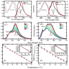
Absorption (dashed) and emission (solid) spectra for (A) PFBT and PFBT-RhB Pdots and (B) PFPV and PFPV-RhB Pdots. The arrow shows absorption peak of RhB in the Pdots. Fluorescence spectra of (C) PFBT-RhB and (D) PFPV-RhB at different temperatures. The ratio of the 573-nm over the 510-nm peaks as a function of temperature were plotted for (E) PFBT-RhB and (F) PFPV-RhB; the insets plot the normalized intensities of the 573-nm peak versus temperature, showing the linear decrease in fluorescence intensity as a function of temperature. The error bars in (E) and (F) represent standard deviation of three independent measurements. To obtain the emission spectra, 450-nm light was used to excite all Pdots.
Figure 3
Figure 3
Confocal microscopy images of HeLa cells labeled with PFBT-RhB at λexc = 458 nm at 13.5°C (A–B) and 36.5°C (D–E); their corresponding bright-field images are shown in (C) and (F). The green channels shown in (A) and (D) were produced by integrating the spectral region from 507 to 518 nm, while the red channels shown in (B) and (E) were from 571 to 582 nm. The scale bar is 20 μm. The arrows in (C) and (F) point to same cell whose integrated spectra were shown in Figure S1 (supporting materials).

References

    1. DeBerardinis RJ, Lum JJ, Hatzivassiliou G, Thompson CB. Cell metabolism. 2008;7:11–20. - PubMed
    1. Borisov S, Klimant I. J Fluor. 2008;18:581–589. - PubMed
    1. Peng HS, Huang SH, Wolfbeis O. J Nanopart Res. 2010;12:2729–2733.
    1. Peng H, Stich MIJ, Yu J, Sun Ln, Fischer LH, Wolfbeis OS. Adv Mater. 2010;22:716–719. - PubMed
    1. Yu J, Sun L, Peng H, Stich MIJ. J Mater Chem. 2010;20:6975–6981.

Publication types