. 2011 Sep;17(17-18):2291-303.
doi: 10.1089/ten.TEA.2010.0682. Epub 2011 Jul 28.

A tissue-engineered muscle repair construct for functional restoration of an irrecoverable muscle injury in a murine model

Affiliations

A tissue-engineered muscle repair construct for functional restoration of an irrecoverable muscle injury in a murine model

Masood A Machingal et al. Tissue Eng Part A. 2011 Sep.

Abstract

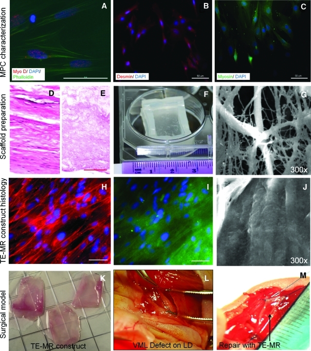
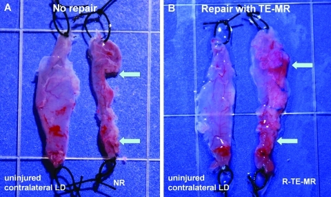
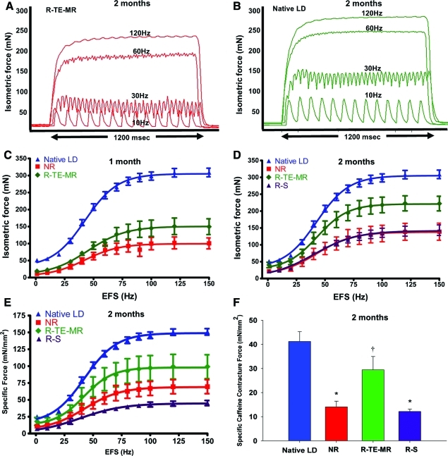
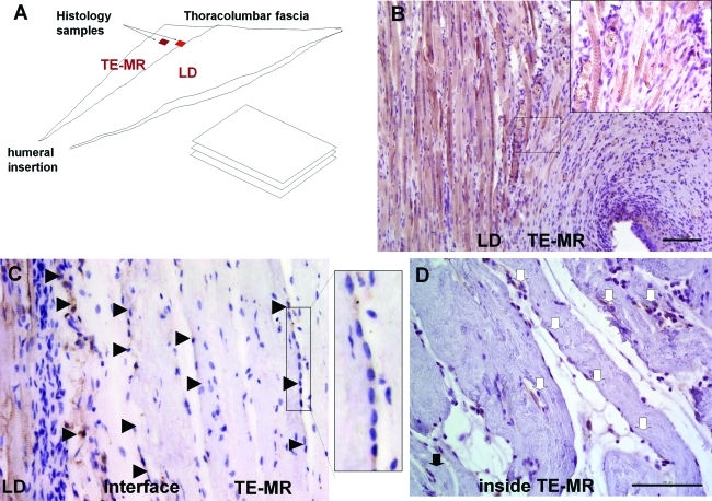
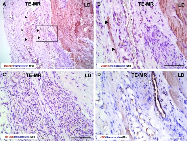
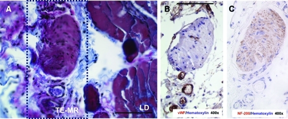
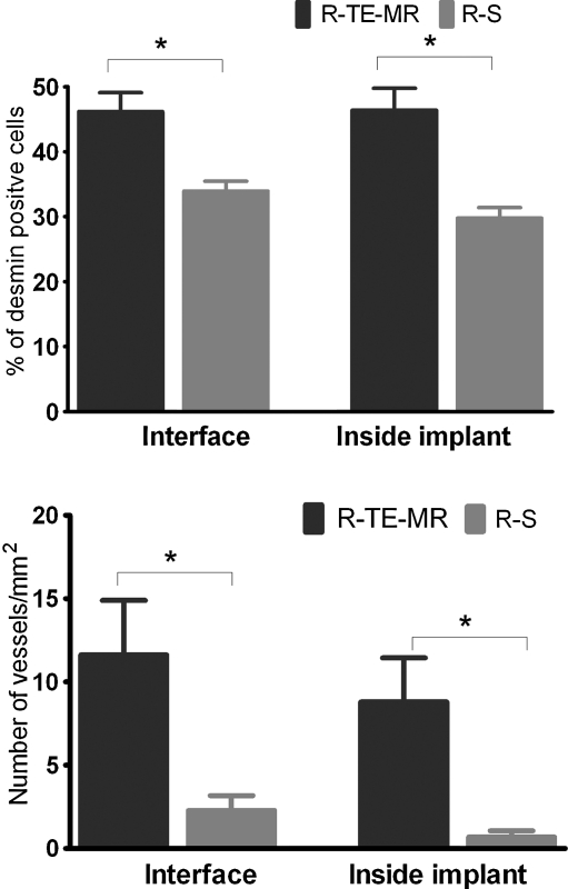
There are no effective clinical treatments for volumetric muscle loss (VML) resulting from traumatic injury, tumor excision, or other degenerative diseases of skeletal muscle. The goal of this study was to develop and characterize a more clinically relevant tissue-engineered muscle repair (TE-MR) construct for functional restoration of a VML injury in the mouse lattissimus dorsi (LD) muscle. To this end, TE-MR constructs developed by seeding rat myoblasts on porcine bladder acellular matrix were preconditioned in a bioreactor for 1 week and implanted in nude mice at the site of a VML injury created by excising 50% of the native LD. Two months postinjury and implantation of TE-MR, maximal tetanic force was ∼72% of that observed in native LD muscle. In contrast, injured LD muscles that were not repaired, or were repaired with scaffold alone, produced only ∼50% of native LD muscle force after 2 months. Histological analyses of LD tissue retrieved 2 months after implantation demonstrated remodeling of the TE-MR construct as well as the presence of desmin-positive myofibers, blood vessels, and neurovascular bundles within the TE-MR construct. Overall, these encouraging initial observations document significant functional recovery within 2 months of implantation of TE-MR constructs and provide clear proof of concept for the applicability of this technology in a murine VML injury model.


Figures

FIG. 1.
FIG. 1.
Development and in vitro assessment of tissue-engineered muscle repair (TE-MR) constructs. Immunofluoresence imaging of muscle progenitor cells (MPCs) (A–C) indicates the presence of muscle-specific markers such as myo-D (A), desmin (B) and myosin (C). Preparation of bladder acellular matrix (BAM) scaffold is shown in (D–F). Representative cross-sectional hematoxylin and eosin images of native porcine bladder (D) before and after and decellularization (E) indicate the absence of cellular materials in the scaffold and preservation of the extracellular matrix (scale bar=100 μm). After the decellularization process, the BAM scaffolds were placed on a custom-made seeding chamber (F) and seeded with MPCs. Scanning electron microscopy imaging was performed to evaluate the cellular coverage on the BAM before (G) and 7 days after static seeding (J) with MPCs. Myotube formation and cellular coverage were observed on ∼95% of the BAM scaffold surface area. Immunostaining with phalloidin (H) and desmin (I) demonstrates the aligned multinucleated (DAPI) myofibers in a bioreactor preconditioned TE-MR construct (scale bar 50 μm). TE-MR constructs after bioreactor preconditioning (K) were implanted in a volumetric muscle loss (VML) injury model in mice. VML injury was developed by excising ∼50% of the native lattissimus dorsi (LD) resulting in a volumetric muscle defect (L, excised area indicated by black dotted lines). The defect was repaired by suturing either TE-MR constructs or scaffolds without cells at the site of excised sites (M; arrow points to implant). Color images available online at www.liebertonline.com/tea
FIG. 2.
FIG. 2.
Morphological assessment of retrieved tissues from the no repair (NR; A) and repair (R-TE-MR; B) treatment groups, compared to the contralateral LD 2 months after implantation. Arrows indicate the original site of the surgical defect. Morphological examination of tissue demonstrates robust tissue formation and remodeling of the TE-MR construct, but little or no tissue formation in the NR group. Color images available online at www.liebertonline.com/tea
FIG. 3.
FIG. 3.
Functional recovery of injured LD after implantation of TE-MR construct. Representative tracings showing the isometric contractions elicited in response to electrical field stimulation (EFS) frequencies of 10, 30, 60, and 120 Hz by R-TE-MR (A) assessed in vitro 2 months after implantation and by uninjured contralateral LD muscle (B). The mean values for the EFS-induced contractions observed on all retrieved tissues in each study group are depicted for both the 1 month (C, n: native LD=20, NR=7, R-TE-MR=6) and 2 month (D, n: native LD=20, NR=5, R-TE-MR=5 and R-S=5) time points, as expressed in both isometric absolute force (mN; C&D), and specific force as a function of stimulation frequency (E, n: native LD=20, NR=5, R-TE-MR=4 and R-S=5). Additionally, after force-frequency testing contralateral native LD muscles (n=6), NR (n=4), R-TE-MR (n=3), or R-S (n=4) at the 2 month time point were subjected to twitch contractions at 0.2 Hz in the presence of a maximally stimulating concentration of caffeine (50 mM) and (F). *Group means are significantly different from that of control (p<0.05). Values are means±standard error of the mean. Group mean is significantly different from that of all other groups (p<0.05). Color images available online at www.liebertonline.com/tea
FIG. 4.
FIG. 4.
Remodeling of TE-MR constructs and muscle tissue formation after implantation in vivo. (A) Schematic diagram of histological sample preparation from the explanted tissue indicating the location of native LD muscle and TE-MR construct, as well as the sample analysis paradigm. Immunohistological staining with desmin shows the presence of striated desmin-positive muscle fibers formed at the LD-TE-MR interface (B). Desmin-positive multinucleated myotubes (black arrowheads) were also observed at the LD-TE-MR interface as well as within the TE-MR construct (∼450–500 μm from interface) (C). Desmin-positive myoblasts (white arrows) were also observed within the construct (D). All scale bars=100 μm. Color images available online at www.liebertonline.com/tea
FIG. 5.
FIG. 5.
Histological analysis of R-TE-MR retrieved 2 months after implantation. Immunohistological staining with desmin (A: 100×and B: 400×) provides further evidence for the presence of myofibers (black arrowheads) and blood vessels (white arrowheads) inside the TE-MR construct. NF-200 staining (C) indicates the presence of nerves at the interface of the native LD muscle and TE-MR construct. Staining with Von Willebrand factor (vWF) (D) demonstrates the presence of blood vessels (white arrowheads) at the interface of the native LD muscle and TE-MR construct. Black rectangle in A represents the approximate location of B and white rectangle represents approximate location of C. All scale bars=100 μm. Color images available online at www.liebertonline.com/tea
FIG. 6.
FIG. 6.
Formation of neurovascular bundles at the interface of native LD of R-TE-MR retrieved 2 months after implantation. Neurovascular bundle stained with herovici polychrome (A), vWF (B) and NF-200 (C) in serial sections. Box in A represents the area of interest for B and C. All scale bars=100 μm. Color images available online at www.liebertonline.com/tea
FIG. 7.
FIG. 7.
Quantification of desmin-positive cells and vessel formation in after repair with TE-MR (R-TE-MR) or scaffold alone (R-S, i.e., scaffold with no cells). As illustrated, desmin-positive staining was significantly (p<0.05) greater in the R-TE-MR group than in the R-S group both at the interface and inside the implanted construct. Formation of blood vessels in the TE-MR was also significantly (p<0.05) greater in R-TE-MR than that of R-S, again, at both the interface and inside the implant; data analyses based on 19 high power field from three different retrieved R-TE-MR tissues and 32 high-power field from four different R-S tissues.

References

    1. Ambrosio F. Kadi F. Lexell J. Fitzgerald G.K. Boninger M.L. Huard J. The effect of muscle loading on skeletal muscle regenerative potential: an update of current research findings relating to aging and neuromuscular pathology. Am J Phys Med Rehabil. 2009;88:145. - PMC - PubMed
    1. Ciciliot S. Schiaffino S. Regeneration of mammalian skeletal muscle. Basic mechanisms and clinical implications. Curr Pharm Des. 2010;16:906. - PubMed
    1. Jarvinen T.A. Jarvinen T.L. Kaariainen M. Kalimo H. Jarvinen M. Muscle injuries: biology and treatment. Am J Sports Med. 2005;33:745. - PubMed
    1. Lee J.Y. Qu-Petersen Z. Cao B. Kimura S. Jankowski R. Cummins J. Usas A. Gates C. Robbins P. Wernig A. Huard J. Clonal isolation of muscle-derived cells capable of enhancing muscle regeneration and bone healing. J Cell Biol. 2000;150:1085. - PMC - PubMed
    1. Moss F.P. Leblond C.P. Nature of dividing nuclei in skeletal muscle of growing rats. J Cell Biol. 1970;44:459. - PMC - PubMed

Publication types